Cell Cycle

Introduction

A cell cycle is a series of events that take place in a cell as it grows and divides. A cell spends most of its time in what is called interphase, and during this time it grows, replicates its chromosomes, and prepares for cell division. The cell then leaves interphase, undergoes mitosis, and completes its division. The resulting cells, known as daughter cells, each enter their own interphase and begin a new round of the cell cycle.

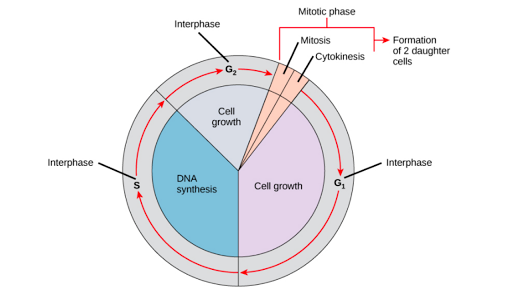
Gentaur Cell Cycle is the ordered sequence of events that occur in a cell in preparation for cell division. The cell cycle is a four-stage process in which the cell increases in size (gap 1, or G1, stage), copies its DNA (synthesis, or S, stage), prepares to divide (gap 2, or G2, stage), and divides (mitosis, or M stage). The G1, S, and G2 stages from interphase represent the time between cell divisions. Based on the stimulating and inhibitory messages a cell receives, it “decides” whether to enter the cell cycle and divide.

Proteins that play a role in stimulating cell division can be classified into four groups: growth factors, growth factor receptors, signal transducers, and nuclear regulatory proteins (transcription factors). For a stimulating signal to reach the nucleus and “activate” cell division, four main steps must occur. First, a growth factor must bind to its receptor on the cell membrane. Second, the receptor must be temporarily activated by this binding event. Third, this activation must stimulate the transmission or transduction of a signal from the receptor on the cell surface to the nucleus within the cell.

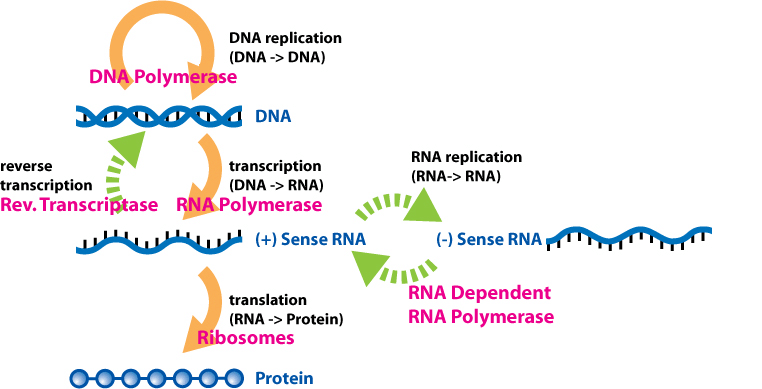
Finally, transcription factors within the nucleus must initiate the transcription of genes involved in cell proliferation. (Transcription is the process by which DNA is converted to RNA. Proteins are then made according to the RNA blueprint, and thus transcription is crucial as the initial step in protein production.) Cells use special proteins and checkpoint signalling systems to ensure that the cell cycle progresses correctly. Checkpoints at the end of G1 and the beginning of G2 are designed to assess DNA damage before and after the S phase. Likewise, a checkpoint during mitosis ensures that the cell’s spindle fibres are properly aligned in metaphase before the chromosomes separate in anaphase.

If DNA damage or spindle formation abnormalities are detected at these checkpoints, the cell is forced to undergo programmed cell death or apoptosis. However, the cell cycle and its checkpoint systems can be sabotaged by defective proteins or genes that cause malignant transformation of the cell, which can lead to cancer. For example, mutations in a protein called p53, which normally detects DNA abnormalities at the G1 checkpoint, may allow cancer-causing mutations to bypass this checkpoint and allow the cell to escape apoptosis.

Cell cycle stages

To divide, a cell must complete several important tasks: It must grow, copy its genetic material (DNA), and physically divide into two daughter cells. Cells perform these tasks in a series of organized and predictable steps that make up the cell cycle. The cell cycle is a cycle, rather than a linear pathway because, at the end of each round, the two daughter cells can start the exact same process from the beginning. In eukaryotic cells or cells with a nucleus, the stages of the cell cycle are divided into two main phases: interphase and the mitotic (M) phase.

  • During interphase, the cell grows and makes a copy of its DNA.
  • During the mitotic (M) phase, the cell separates its DNA into two sets and divides its cytoplasm, forming two new cells.

M phase

During the mitotic (M) phase, the cell splits its copied DNA and cytoplasm to form two new cells. The M phase involves two distinct processes related to division: mitosis and cytokinesis.

In mitosis, the cell’s nuclear DNA condenses into visible chromosomes and is pulled apart by the mitotic spindle, a specialized structure made of microtubules. Mitosis occurs in four stages: prophase (sometimes divided into early prophase and prometaphase), metaphase, anaphase, and telophase. You can learn more about these stages in the video on mitosis.

In cytokinesis, the cytoplasm of the cell splits in two, forming two new cells. Cytokinesis usually begins just as mitosis ends, with a little overlap. Importantly, cytokinesis is carried out differently in animal and plant cells.
Cytokinesis in animal and plant cells.

  • In an animal cell, a contractile ring of cytoskeletal fibres forms in the middle of the cell and contracts inward, producing a cleft called the cleavage furrow. Eventually, the contractile ring pinches the parent cell in two, producing two daughter cells.
  • In a plant cell, vesicles derived from the Golgi apparatus move toward the centre of the cell, where they fuse to form a structure called a cell plate. The cell plate expands outward and connects with the cell’s lateral walls, creating a new cell wall that divides the parent cell to form two daughter cells.

cDNA (Complementary DNA)

(cDNA) Complementary DNA is a double-stranded DNA version of an mRNA molecule. In higher eukaryotes, an mRNA is a more useful predictor of a polypeptide sequence than a genomic sequence, because the introns have been separated. Researchers prefer to use cDNA over mRNA because RNAs are inherently less stable than DNA and techniques to routinely amplify and purify individual RNA molecules do not exist.

cDNA is made from mRNA with the use of a special enzyme called reverse transcriptase, originally isolated from retroviruses. Using an mRNA molecule as a template, reverse transcriptase synthesizes a single-stranded DNA molecule that can then be used as a template for double-stranded DNA synthesis. It is not necessary to cut the cDNA in order to clone it.

DNA: the building block of life

Deoxyribonucleic acid (DNA) is the molecule that carries the instructions for all aspects of an organism’s functions, from growth to metabolism to reproduction. In living organisms, most of the DNA resides in tightly coiled structures called chromosomes, located within the nucleus of each cell. DNA is made up of four different building blocks, called nucleotides, each of which is made up of one of the four nitrogenous bases. These are the purines: guanine (G) and adenine (A), and the pyrimidines: thymine (T) and cytosine (C).

These nucleotides are attached to a deoxyribose sugar and can join other deoxyribose sugars through phosphate bonds to form long chains, some of which can be more than 100,000,000 molecules long. Since each deoxyribose in a DNA strand is attached to one of four nitrogenous bases (G, A, T, or C), these long chains can carry information.

Groups of three nucleotides form the smallest but most well-defined “words” in the language of DNA. These “words” are called codons. Codons are used to request the joining of specific amino acids to form proteins. For example, the adenosine-adenosine-guanosine (AAG) codon requires the amino acid lysine (Lys) to be incorporated into a protein molecule. The AGG codon calls the amino acid arginine (arg). So AAG-AGG would require a lys to be coupled to an arg in a growing protein chain. There are also codons that, under the right circumstances, require a protein to begin to form (start codons) or a protein chain to end (stop codons). As you can see from this simple example, DNA can carry a huge amount of information.

What is genomic and complementary DNA?

The DNA that resides on the chromosomes inside the nucleus, with all the biological information that will be transferred to the next generation, is called genomic DNA (gDNA). The words “genome” and “genomic” come from the word “gene”. A gene is a set of codons that specify a specific protein chain, along with associated start and stop codons. The word genome is an extension of this concept and means the collection of all the genes and other information contained within the nuclei of the cells of an organism. Often when the word “DNA” is used without further clarification, it refers to gDNA.

In nature, the process for information to be transmitted from DNA can occur through gene replication or gene expression. There are some important factors to keep in mind:

  • DNA can copy itself in a process known as replication, using DNA polymerase.
  • Information from DNA passes through messenger RNA (mRNA), which contains sets of four nucleotides (uracil, adenine, guanine, and cytosine).
  • mRNA is produced when enzymes, such as RNA polymerase, bind to specific genes and copy their information into RNA using ribose sugar (not deoxyribose as in DNA). This process is called transcription.
  • Ribosomes assemble around the mRNA, creating a chain of amino acids to create specific proteins. This is called translation.
  • Due to the ribose sugar chains, mRNA is short-lived. It is designed to transmit information from the chromosomes in the nucleus to the machinery that makes proteins.
  • The mRNA rapidly degrades after it has completed its purpose.

Initially, it was observed that gDNA was always read and transcribed into mRNA, which guided protein formation, and was then removed. The notion that information can always flow from DNA to RNA to protein was jokingly called the central dogma of molecular biology.

The functions of gDNA and cDNA

cDNA can be described as gDNA without all the necessary non-coding regions, thus it gets its name as complementary DNA. The main distinction to be made between cDNA and gDNA is the existence of introns and exons. Introns are nucleotides in genes that do not have coding sequences. Introns are usually cleaved or “removed” from RNA in the transcription process before proteins are created. It should be noted that prokaryotes are not capable of splicing introns. Exons are a necessary part of the coding system and are retained after introns are spliced. Extron’s are non-splicing introns, even though they do not contain coding sequences.

When scientists use viral enzymes to make cDNA from RNA isolated from the cells and tissues they are studying, it does not contain introns because it is spliced ​​into mRNA. The cDNA also does not contain any other gDNA that does not directly code for a protein (referred to as non-coding DNA). Lastly, not all genes in gDNA are transcribed into mRNA at any given time. As a result, the cDNA will only contain genes that are actively being used by a specific cell or tissue at any given time. There is much less total information in cDNA than in gDNA, but the information that remains may be much more relevant to what a researcher is looking for since it does not contain sequences that are unnecessary for DNA function and replication.

Once isolated, gDNA can be used to create genomic libraries for DNA sequencing, fingerprinting, differentiation, and other applications in both the clinical and research fields. cDNA can also be used to make cDNA libraries, permanent collections of cDNA that can be copied and/or stored long-term and is commonly used to clone eukaryotic genes into a prokaryote. In this way, a protein expressed in a eukaryotic organism can be introduced into a prokaryote. For this process, cDNA over gDNA is used, since prokaryotes cannot stimulate introns contained in gDNA.

To isolate cDNA, RNA must first be isolated from an organism. Then, using a reverse transcriptase enzyme, cDNA can be produced. This is the process that retroviruses use to incorporate themselves into the cells of their host. Retroviruses, such as simian immunodeficiency virus (SIV) and avian myeloblastosis virus (AMV), use their cDNA to produce mRNA in the host, leading to the production of viral proteins. This is possible because retroviruses use RNA as genomic material instead of DNA, and it is reverse transcribed into cDNA, which then undergoes normal transcription and leads to viral protein in the host.

Custom and pre-made gDNA and cDNA available on BioChain

BioChain provides access to a comprehensive and well-documented tissue bank containing isolated samples that have been tested for contaminants. As part of rigorous quality control, gDNA samples are analyzed by spectrophotometer and electrophoresis, with concentration determined by UV260 measurement and plant concentration determined by green peak measurement. All gDNA is treated with RNase to remove all RNA.

Genomic DNA comes from unique sources, including hundreds of healthy or diseased organ tissues from humans, animals, and plants. gDNA has applications ranging from SNP analysis, methylation studies, copy number variation (CNV) analysis, comparative genomic hybridization (CGH), Southern blotting, next-generation sequencing, and PCR.

Tryptophan Repressor

Abstract

Tryptophan biosynthesis in Escherichia coli is regulated by the trpR gene product, the Gentaur Tryptophan Repressor (Trp). The trp aporepressor binds to the corepressor, L-tryptophan, to form a holopressor complex, which binds tightly to the trp operator DNA and inhibits transcription of the tryptophan biosynthetic operon. The conservation of trp operator sequences among enteric Gram-negative bacteria suggests that trpR genes from other bacterial species can be cloned by complementation in E. coli. To clone trpR homologues, the E. coli trpR gene, delta trpR504, was deleted from a plasmid by site-directed mutagenesis, then crossed into the E. coli genome.

Plasmid clones of Enterobacter aerogenes and Enterobacter cloacae trpR genes were isolated by complementation of the trpR504 delta allele, qualified as the ability to repress beta-galactosidase synthesis from a prophage-transmitted trpE-lacZ gene fusion. The predicted amino acid sequences of four enteric TrpR proteins show differences, clustered at the back of the folded repressor, versus DNA-binding helix-turn-helix substructures.

These differences are predicted to have little effect on interactions of aporepressor with tryptophan, holorepressor with operator DNA, or holorepressor dimers linked in tandem with each other. Although some variation in the dimer interface is observed, the interactions expected to stabilize the interface are preserved. The phylogenetic relationships revealed by the TrpR amino acid sequence alignment are consistent with the results of others.

In E. coli, the synthesis of the amino acid tryptophan from precursors available to the cell requires 5 enzymes. The genes that encode them are grouped into a single operon with its own promoter and operator. When tryptophan is available to the cell, its presence turns off the operon.

Mechanism

  • One tryptophan molecule binds to one site on each Trp repressor monomer.
  • The Trp repressor, a homodimer of two of these complexes, binds to the operator of the Trp operon.
  • This stops the transcription of all 5 genes in the operon, so the enzymes used in Trp synthesis are not synthesized.

This stereoscopic view () shows the tryptophan repressor (right side of each panel) bound to its operator DNA (left side). The two identical repressor polypeptides are shown on either side of the horizontal red line. The two tryptophan molecules are shown as red rings. Also, look for the alpha-helix stretches in each monomer. You may find it easier to merge the two images into a 3D view by holding an 8.5 x 11″ (22 x 28 cm) sheet of paper vertically between your nose and the dividing line between the two images on the screen so that your left eye sees only the image on the left, his right eye only the right.