Rubella Spike Ectodomain (E1-E2) Antigen: Structure, Function, and Applications

Rubella, a mild yet highly contagious viral illness caused by the Rubella virus (RuV), has significant implications for public health, particularly due to its association with congenital rubella syndrome (CRS). CRS can result in severe birth defects, including cardiac abnormalities, cataracts, and developmental delays. The Rubella Spike Ectodomain (E1-E2) Antigen, consisting of two glycoproteins (E1 and E2), has emerged as a focal point for advancing diagnostics, vaccine development, and therapeutic strategies.

This article explores the structural and functional attributes of the E1-E2 ectodomain and its critical role in rubella research and public health initiatives.
Molecular and Structural Insights into the E1-E2 Antigen

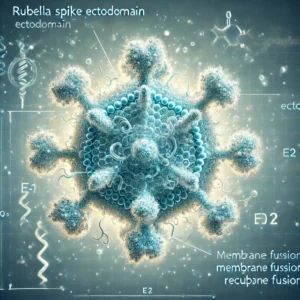
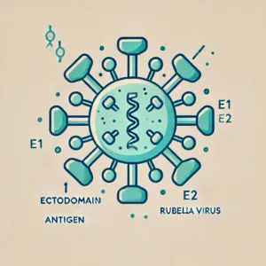
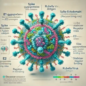
The spike ectodomain of RuV is primarily composed of two glycoproteins:

  • E1 Glycoprotein: A class II fusion protein responsible for viral entry into host cells.
  • E2 Glycoprotein: Stabilizes the E1 trimer and mediates receptor interactions critical for cell attachment.

E1 Glycoprotein: The Fusion Catalyst

The E1 glycoprotein drives the viral membrane fusion process, facilitating the injection of viral RNA into the host cytoplasm. Studies on the structural mechanisms of E1, published by NIH, demonstrate that specific regions within E1 are highly conserved, making it a prime target for neutralizing antibodies. The Protein Data Bank provides a repository of high-resolution structures that have been instrumental in understanding these functional domains.

E2 Glycoprotein: Enhancing Viral Stability

The E2 glycoprotein works in concert with E1, enhancing stability and ensuring proper viral attachment. Research from the CDC has highlighted the immunological relevance of E2 in triggering T-cell-mediated responses. Studies from Johns Hopkins University further underscore its role in modulating host immune evasion.

The combination of these glycoproteins in the E1-E2 ectodomain forms a critical target for vaccine development and diagnostics.

Role in Diagnostic Applications

The E1-E2 antigen is central to diagnostic tools for detecting RuV-specific antibodies. Its recombinant form has enabled the development of high-sensitivity ELISA kits. These kits are approved by regulatory agencies such as the FDA and recommended by the World Health Organization (WHO) for confirming rubella immunity, especially in prenatal care.

Advanced Diagnostics

  1. ELISA Tests: Incorporation of recombinant E1-E2 enhances test accuracy, reducing false positives. These assays, as reported by Stanford University, offer unparalleled sensitivity in detecting IgG and IgM antibodies.
  2. PCR-Based Detection: Quantitative PCR assays targeting E1 gene sequences provide rapid and reliable rubella diagnosis. The National Institute of Allergy and Infectious Diseases (NIAID) highlights their application in outbreak scenarios.
  3. Point-of-Care Devices: Portable diagnostic tools utilizing E1-E2 antigens, supported by Harvard University, are being deployed in low-resource settings to improve early detection and intervention.
Significance in Vaccine Development

Rubella vaccines are a cornerstone of global immunization programs, reducing rubella incidence and preventing CRS. Traditional live-attenuated vaccines include the full viral particle, but there is growing interest in antigen-specific subunit vaccines based on the E1-E2 ectodomain.

Subunit Vaccines Using E1-E2

  • Advantages: Subunit vaccines minimize the risks associated with live attenuated strains, especially in immunocompromised individuals. They also provide opportunities for targeting specific immune responses, as documented in research from Oxford University.
  • Recombinant Platforms: Studies published by MIT demonstrate how recombinant E1-E2 antigens expressed in Escherichia coli or insect cell systems enhance vaccine immunogenicity.

Research from the NIH Vaccine Research Center is actively investigating these next-generation vaccines, which are poised to meet the goals of the Global Rubella Elimination Initiative led by WHO.

Therapeutic Potential and Monoclonal Antibodies

The development of therapeutic monoclonal antibodies targeting the E1-E2 ectodomain is another promising avenue. High-affinity monoclonal antibodies are being tested for their ability to neutralize RuV and prevent severe outcomes in CRS. Data from the Mayo Clinic show encouraging preclinical results.

Monoclonal Antibody Development

  • Neutralizing E1 Antibodies: Studies hosted by PubMed indicate that E1-specific antibodies can block viral entry effectively.
  • Combination Therapies: Combining E1 and E2 targeting antibodies is under investigation at Yale University.
Global Implications and Future Directions

Rubella continues to challenge public health systems, particularly in regions where vaccination rates remain low. The E1-E2 spike ectodomain is at the forefront of efforts to combat this disease through innovative diagnostics, vaccines, and therapeutics.

Future Research Directions

  1. Structural Studies: Further investigation into the E1-E2 interaction using cryo-electron microscopy, as detailed by Nature, will provide deeper insights into therapeutic targeting.
  2. CRS Prevention: Collaborative research funded by Bill & Melinda Gates Foundation aims to optimize the use of E1-E2 antigens in vertical transmission prevention strategies.
  3. Immune Escape Mechanisms: Research at UC Berkeley is exploring mutations in the E1-E2 region that enable immune evasion, informing vaccine redesign.

Conclusion

The Rubella Spike Ectodomain (E1-E2) Antigen represents a crucial element in the fight against rubella. Its structural and immunological significance has enabled advancements in diagnostics, vaccine development, and therapeutic approaches. As global health organizations like the WHO and CDC work towards rubella elimination, the E1-E2 ectodomain remains central to these efforts.

For additional resources and research, visit trusted repositories like NIH, PubMed, and academic institutions such as Harvard and Stanford.

By leveraging the power of the E1-E2 antigen, the global medical community is poised to achieve significant milestones in rubella control and prevention.

AIF1 / Iba1 (Microglia Marker) CF647 Conjugate Antibody: Advanced Tool for Microglial Research

The AIF1/Iba1 (Microglia Marker) CF647 Conjugate Antibody is a specialized fluorescent antibody targeting Allograft Inflammatory Factor 1 (AIF1), also known as Iba1. This protein is a critical marker for microglia, the immune cells of the central nervous system (CNS), and plays a key role in neuroinflammation, neurodegeneration, and CNS homeostasis. The CF647 conjugation provides superior fluorescence properties for imaging and flow cytometry, making it an invaluable tool for advanced neurological research.

Overview of AIF1/Iba1

Key Features:

  • Full Name: Allograft Inflammatory Factor 1 (AIF1), also known as Ionized Calcium-Binding Adaptor Molecule 1 (Iba1).
  • Function:
    • Regulates microglial activation.
    • Involved in cytoskeletal reorganization.
    • Facilitates phagocytosis and immune responses in the CNS.
  • Clinical Significance:
    • Highly expressed in activated microglia during neuroinflammatory and neurodegenerative conditions, such as Alzheimer’s disease, Parkinson’s disease, and multiple sclerosis.

For more information, visit NCBI Gene Database.

Features of the AIF1/Iba1 CF647 Conjugate Antibody

  1. Fluorescence Conjugation:
    • Conjugated to CF647, a bright, photostable far-red fluorophore for superior imaging performance.
    • Compatible with standard confocal and fluorescence microscopy filters.
  2. High Specificity:
    • Targets AIF1/Iba1 with minimal cross-reactivity, ensuring precise identification of microglia.
  3. Versatility:
    • Suitable for:
      • Immunofluorescence (IF): Visualizes microglia in tissue sections.
      • Flow Cytometry (FC): Quantifies microglial populations in cell suspensions.
  4. Species Reactivity:
    • Validated for human, mouse, and rat samples.
  5. Ready-to-Use:
    • Supplied at 0.1 mg/mL for immediate use in standard protocols.

Applications in Research

  1. Neuroinflammation Studies:
    • Tracks microglial activation in response to injury or disease.
  2. Neurodegenerative Diseases:
    • Examines microglial involvement in Alzheimer’s, Parkinson’s, and Huntington’s disease.
  3. Immune Responses in CNS:
    • Studies the role of microglia in autoimmune disorders like multiple sclerosis.
  4. Drug Development:
    • Evaluates therapeutic compounds targeting microglial activation and neuroinflammation.

Protocols for Using AIF1/Iba1 CF647 Antibody

Immunofluorescence (IF):

  1. Sample Preparation:
    • Fix tissue sections with 4% paraformaldehyde.
    • Permeabilize with 0.1% Triton X-100.
  2. Antibody Incubation:
    • Dilute CF647 conjugated antibody (1:200–1:500 recommended).
    • Incubate for 1 hour at room temperature in the dark.
  3. Imaging:
    • Use a fluorescence microscope with appropriate filters for CF647.

Flow Cytometry (FC):

  1. Cell Preparation:
    • Isolate microglial cells from CNS tissue using enzymatic digestion and gradient separation.
    • Fix and permeabilize cells as needed.
  2. Staining:
    • Incubate cells with the antibody diluted in buffer (1:50–1:200).
    • Wash and resuspend in flow cytometry buffer.
  3. Detection:
    • Use a flow cytometer with far-red laser compatibility.

Benefits of the AIF1/Iba1 CF647 Antibody

  1. High Sensitivity and Specificity:
    • Accurately labels microglia in CNS tissues.
  2. Superior Fluorescence:
    • CF647 offers brighter signals and reduced photobleaching compared to other dyes.
  3. Multifunctional:
    • Suitable for imaging, flow cytometry, and co-localization studies.
  4. Reproducibility:
    • Consistent results across various experimental setups.

Challenges and Future Directions

Challenges:

  • Fluorophore Compatibility:
    • Requires compatible microscopy and cytometry equipment.
  • Optimization:
    • Staining conditions may need adjustment for different sample types.

Future Innovations:

  1. Multiplex Antibody Panels:
    • Combine with antibodies targeting other microglial markers for comprehensive profiling.
  2. Super-Resolution Imaging:
    • Leverage CF647 for high-resolution studies of microglial dynamics.
  3. Therapeutic Applications:
    • Utilize AIF1/Iba1 as a biomarker for microglial-targeted therapies.

Conclusion

The AIF1/Iba1 CF647 Conjugate Antibody is an advanced tool for exploring microglial biology and CNS immunity. Its high specificity, versatile applications, and superior fluorescence properties make it indispensable for cutting-edge research in neuroscience and immunology.

For further resources and technical support, visit:

Caltrin ELISA: A Key Tool in Protein Detection and Research

Caltrin (Calcium Transport Inhibitor) is a protein that plays a crucial role in regulating calcium ion channels, specifically in the reproductive system. The Caltrin ELISA (Enzyme-Linked Immunosorbent Assay) is a specialized assay designed to detect and quantify Caltrin protein levels in biological samples. This assay is widely used in reproductive biology, molecular diagnostics, and protein research.

What is Caltrin?

Caltrin is a small protein primarily found in the male reproductive system. It binds to spermatozoa and inhibits calcium uptake, preventing premature activation of sperm cells before they enter the female reproductive tract. Caltrin proteins play a crucial role in sperm viability and functionality, which makes them important targets for research on fertility and reproductive health.

The ability to accurately measure Caltrin levels in biological samples, such as semen or reproductive tissues, is essential for understanding its role in sperm physiology and for diagnosing conditions related to male fertility.

How Caltrin ELISA Works

The Caltrin ELISA is based on the principles of enzyme-linked immunosorbent assays, which involve the detection of a specific antigen (Caltrin protein) using antibodies. The assay typically follows these steps:

  1. Capture Antibody: A specific antibody against Caltrin is coated onto a microplate.
  2. Sample Application: Biological samples, such as semen, are added to the wells, where Caltrin binds to the antibody.
  3. Detection Antibody: A second antibody, labeled with an enzyme, binds to the captured Caltrin protein.
  4. Substrate Addition: A substrate for the enzyme is added, producing a detectable signal, usually a color change, which correlates with the amount of Caltrin in the sample.

The intensity of the color can be measured using a spectrophotometer, allowing researchers to quantify the concentration of Caltrin in the sample.

Applications of Caltrin ELISA

  1. Fertility Research: Caltrin is a critical player in sperm function, particularly in regulating calcium channels that are essential for sperm motility and viability. According to NIH, the Caltrin ELISA is valuable for assessing male fertility and diagnosing conditions that affect sperm function.
  2. Reproductive Health Diagnostics: Measuring Caltrin levels can provide insights into certain reproductive disorders. For instance, reduced levels of Caltrin could indicate sperm dysfunction, contributing to infertility. The Centers for Disease Control and Prevention (CDC) emphasizes the importance of protein-based assays in diagnosing infertility issues.
  3. Semen Analysis: In clinical settings, Caltrin ELISA can be used alongside other semen analysis tools to provide a more comprehensive picture of sperm health. Studies published by PubMed Central (PMC) show that Caltrin levels can be correlated with sperm motility, offering a potential marker for reproductive success.
  4. Calcium Channel Research: Since Caltrin is involved in the inhibition of calcium ion transport in sperm cells, its quantification is vital for understanding calcium signaling pathways. The FDA and NIH note that calcium transport proteins like Caltrin are critical in cellular functions, making their study important for broader biological research.

Advantages of Using Caltrin ELISA

  1. High Sensitivity and Specificity: The Caltrin ELISA is designed to specifically detect and quantify Caltrin protein, offering high sensitivity in even low-abundance samples. This precision is critical for reliable reproductive research, as noted by Johns Hopkins University in their studies on fertility markers.
  2. Quantitative Analysis: Unlike qualitative tests, the Caltrin ELISA provides a quantitative measurement of protein concentration, allowing researchers to detect small changes in protein levels that may be indicative of physiological or pathological conditions.
  3. Non-Invasive Sampling: The assay requires small sample volumes, typically using semen or other reproductive tissues. This minimizes the invasiveness of the procedure, making it a suitable option for both clinical and research applications.
  4. Consistency and Reproducibility: Caltrin ELISA kits are produced under strict quality control guidelines, such as GMP standards, ensuring reproducible results across different batches and experiments.

Diagnostic Potential and Clinical Impact

The Caltrin ELISA offers diagnostic potential in male fertility disorders. Reduced levels of Caltrin protein could be linked to conditions like asthenozoospermia (reduced sperm motility), one of the leading causes of male infertility. By quantifying Caltrin levels, clinicians can gain insights into sperm health and functionality, guiding treatment decisions for patients undergoing fertility evaluations.

Additionally, understanding Caltrin’s role in calcium transport inhibition may pave the way for novel therapeutic approaches in reproductive medicine. The National Institutes of Health (NIH) and the CDC emphasize the need for reliable biomarkers like Caltrin to improve diagnosis and treatment strategies for infertility.

Conclusion

The Caltrin ELISA is a powerful tool in the field of reproductive biology and diagnostics. By offering precise and reliable measurements of Caltrin protein, this assay contributes significantly to our understanding of sperm physiology and male fertility. Its applications in diagnostics, research, and therapeutic development make it an indispensable tool for reproductive health specialists and molecular biologists alike.

By following GMP guidelines and adhering to rigorous quality control, Caltrin ELISA kits ensure consistent and accurate results, fostering advancements in fertility research and clinical care.

Cell Cycle

Introduction

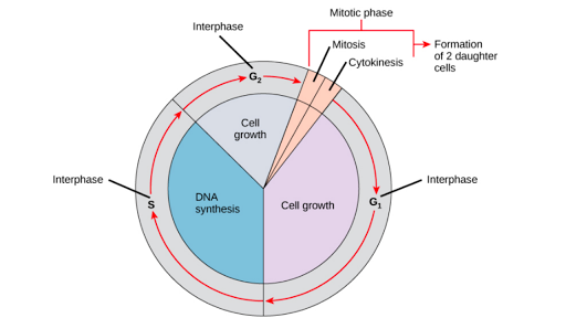
A cell cycle is a series of events that take place in a cell as it grows and divides. A cell spends most of its time in what is called interphase, and during this time it grows, replicates its chromosomes, and prepares for cell division. The cell then leaves interphase, undergoes mitosis, and completes its division. The resulting cells, known as daughter cells, each enter their own interphase and begin a new round of the cell cycle.

Gentaur Cell Cycle is the ordered sequence of events that occur in a cell in preparation for cell division. The cell cycle is a four-stage process in which the cell increases in size (gap 1, or G1, stage), copies its DNA (synthesis, or S, stage), prepares to divide (gap 2, or G2, stage), and divides (mitosis, or M stage). The G1, S, and G2 stages from interphase represent the time between cell divisions. Based on the stimulating and inhibitory messages a cell receives, it “decides” whether to enter the cell cycle and divide.

Proteins that play a role in stimulating cell division can be classified into four groups: growth factors, growth factor receptors, signal transducers, and nuclear regulatory proteins (transcription factors). For a stimulating signal to reach the nucleus and “activate” cell division, four main steps must occur. First, a growth factor must bind to its receptor on the cell membrane. Second, the receptor must be temporarily activated by this binding event. Third, this activation must stimulate the transmission or transduction of a signal from the receptor on the cell surface to the nucleus within the cell.

Finally, transcription factors within the nucleus must initiate the transcription of genes involved in cell proliferation. (Transcription is the process by which DNA is converted to RNA. Proteins are then made according to the RNA blueprint, and thus transcription is crucial as the initial step in protein production.) Cells use special proteins and checkpoint signalling systems to ensure that the cell cycle progresses correctly. Checkpoints at the end of G1 and the beginning of G2 are designed to assess DNA damage before and after the S phase. Likewise, a checkpoint during mitosis ensures that the cell’s spindle fibres are properly aligned in metaphase before the chromosomes separate in anaphase.

If DNA damage or spindle formation abnormalities are detected at these checkpoints, the cell is forced to undergo programmed cell death or apoptosis. However, the cell cycle and its checkpoint systems can be sabotaged by defective proteins or genes that cause malignant transformation of the cell, which can lead to cancer. For example, mutations in a protein called p53, which normally detects DNA abnormalities at the G1 checkpoint, may allow cancer-causing mutations to bypass this checkpoint and allow the cell to escape apoptosis.

Cell cycle stages

To divide, a cell must complete several important tasks: It must grow, copy its genetic material (DNA), and physically divide into two daughter cells. Cells perform these tasks in a series of organized and predictable steps that make up the cell cycle. The cell cycle is a cycle, rather than a linear pathway because, at the end of each round, the two daughter cells can start the exact same process from the beginning. In eukaryotic cells or cells with a nucleus, the stages of the cell cycle are divided into two main phases: interphase and the mitotic (M) phase.

  • During interphase, the cell grows and makes a copy of its DNA.
  • During the mitotic (M) phase, the cell separates its DNA into two sets and divides its cytoplasm, forming two new cells.

M phase

During the mitotic (M) phase, the cell splits its copied DNA and cytoplasm to form two new cells. The M phase involves two distinct processes related to division: mitosis and cytokinesis.

In mitosis, the cell’s nuclear DNA condenses into visible chromosomes and is pulled apart by the mitotic spindle, a specialized structure made of microtubules. Mitosis occurs in four stages: prophase (sometimes divided into early prophase and prometaphase), metaphase, anaphase, and telophase. You can learn more about these stages in the video on mitosis.

In cytokinesis, the cytoplasm of the cell splits in two, forming two new cells. Cytokinesis usually begins just as mitosis ends, with a little overlap. Importantly, cytokinesis is carried out differently in animal and plant cells.
Cytokinesis in animal and plant cells.

  • In an animal cell, a contractile ring of cytoskeletal fibres forms in the middle of the cell and contracts inward, producing a cleft called the cleavage furrow. Eventually, the contractile ring pinches the parent cell in two, producing two daughter cells.
  • In a plant cell, vesicles derived from the Golgi apparatus move toward the centre of the cell, where they fuse to form a structure called a cell plate. The cell plate expands outward and connects with the cell’s lateral walls, creating a new cell wall that divides the parent cell to form two daughter cells.

cDNA (Complementary DNA)

(cDNA) Complementary DNA is a double-stranded DNA version of an mRNA molecule. In higher eukaryotes, an mRNA is a more useful predictor of a polypeptide sequence than a genomic sequence, because the introns have been separated. Researchers prefer to use cDNA over mRNA because RNAs are inherently less stable than DNA and techniques to routinely amplify and purify individual RNA molecules do not exist.

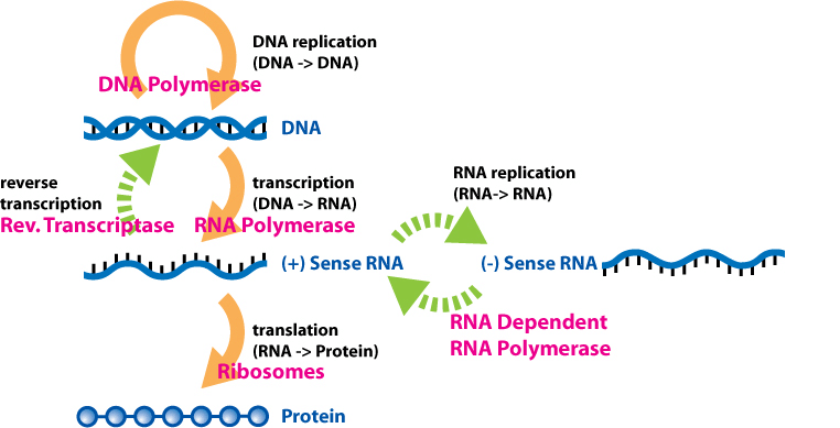
cDNA is made from mRNA with the use of a special enzyme called reverse transcriptase, originally isolated from retroviruses. Using an mRNA molecule as a template, reverse transcriptase synthesizes a single-stranded DNA molecule that can then be used as a template for double-stranded DNA synthesis. It is not necessary to cut the cDNA in order to clone it.

DNA: the building block of life

Deoxyribonucleic acid (DNA) is the molecule that carries the instructions for all aspects of an organism’s functions, from growth to metabolism to reproduction. In living organisms, most of the DNA resides in tightly coiled structures called chromosomes, located within the nucleus of each cell. DNA is made up of four different building blocks, called nucleotides, each of which is made up of one of the four nitrogenous bases. These are the purines: guanine (G) and adenine (A), and the pyrimidines: thymine (T) and cytosine (C).

These nucleotides are attached to a deoxyribose sugar and can join other deoxyribose sugars through phosphate bonds to form long chains, some of which can be more than 100,000,000 molecules long. Since each deoxyribose in a DNA strand is attached to one of four nitrogenous bases (G, A, T, or C), these long chains can carry information.

Groups of three nucleotides form the smallest but most well-defined “words” in the language of DNA. These “words” are called codons. Codons are used to request the joining of specific amino acids to form proteins. For example, the adenosine-adenosine-guanosine (AAG) codon requires the amino acid lysine (Lys) to be incorporated into a protein molecule. The AGG codon calls the amino acid arginine (arg). So AAG-AGG would require a lys to be coupled to an arg in a growing protein chain. There are also codons that, under the right circumstances, require a protein to begin to form (start codons) or a protein chain to end (stop codons). As you can see from this simple example, DNA can carry a huge amount of information.

What is genomic and complementary DNA?

The DNA that resides on the chromosomes inside the nucleus, with all the biological information that will be transferred to the next generation, is called genomic DNA (gDNA). The words “genome” and “genomic” come from the word “gene”. A gene is a set of codons that specify a specific protein chain, along with associated start and stop codons. The word genome is an extension of this concept and means the collection of all the genes and other information contained within the nuclei of the cells of an organism. Often when the word “DNA” is used without further clarification, it refers to gDNA.

In nature, the process for information to be transmitted from DNA can occur through gene replication or gene expression. There are some important factors to keep in mind:

  • DNA can copy itself in a process known as replication, using DNA polymerase.
  • Information from DNA passes through messenger RNA (mRNA), which contains sets of four nucleotides (uracil, adenine, guanine, and cytosine).
  • mRNA is produced when enzymes, such as RNA polymerase, bind to specific genes and copy their information into RNA using ribose sugar (not deoxyribose as in DNA). This process is called transcription.
  • Ribosomes assemble around the mRNA, creating a chain of amino acids to create specific proteins. This is called translation.
  • Due to the ribose sugar chains, mRNA is short-lived. It is designed to transmit information from the chromosomes in the nucleus to the machinery that makes proteins.
  • The mRNA rapidly degrades after it has completed its purpose.

Initially, it was observed that gDNA was always read and transcribed into mRNA, which guided protein formation, and was then removed. The notion that information can always flow from DNA to RNA to protein was jokingly called the central dogma of molecular biology.

The functions of gDNA and cDNA

cDNA can be described as gDNA without all the necessary non-coding regions, thus it gets its name as complementary DNA. The main distinction to be made between cDNA and gDNA is the existence of introns and exons. Introns are nucleotides in genes that do not have coding sequences. Introns are usually cleaved or “removed” from RNA in the transcription process before proteins are created. It should be noted that prokaryotes are not capable of splicing introns. Exons are a necessary part of the coding system and are retained after introns are spliced. Extron’s are non-splicing introns, even though they do not contain coding sequences.

When scientists use viral enzymes to make cDNA from RNA isolated from the cells and tissues they are studying, it does not contain introns because it is spliced ​​into mRNA. The cDNA also does not contain any other gDNA that does not directly code for a protein (referred to as non-coding DNA). Lastly, not all genes in gDNA are transcribed into mRNA at any given time. As a result, the cDNA will only contain genes that are actively being used by a specific cell or tissue at any given time. There is much less total information in cDNA than in gDNA, but the information that remains may be much more relevant to what a researcher is looking for since it does not contain sequences that are unnecessary for DNA function and replication.

Once isolated, gDNA can be used to create genomic libraries for DNA sequencing, fingerprinting, differentiation, and other applications in both the clinical and research fields. cDNA can also be used to make cDNA libraries, permanent collections of cDNA that can be copied and/or stored long-term and is commonly used to clone eukaryotic genes into a prokaryote. In this way, a protein expressed in a eukaryotic organism can be introduced into a prokaryote. For this process, cDNA over gDNA is used, since prokaryotes cannot stimulate introns contained in gDNA.

To isolate cDNA, RNA must first be isolated from an organism. Then, using a reverse transcriptase enzyme, cDNA can be produced. This is the process that retroviruses use to incorporate themselves into the cells of their host. Retroviruses, such as simian immunodeficiency virus (SIV) and avian myeloblastosis virus (AMV), use their cDNA to produce mRNA in the host, leading to the production of viral proteins. This is possible because retroviruses use RNA as genomic material instead of DNA, and it is reverse transcribed into cDNA, which then undergoes normal transcription and leads to viral protein in the host.

Custom and pre-made gDNA and cDNA available on BioChain

BioChain provides access to a comprehensive and well-documented tissue bank containing isolated samples that have been tested for contaminants. As part of rigorous quality control, gDNA samples are analyzed by spectrophotometer and electrophoresis, with concentration determined by UV260 measurement and plant concentration determined by green peak measurement. All gDNA is treated with RNase to remove all RNA.

Genomic DNA comes from unique sources, including hundreds of healthy or diseased organ tissues from humans, animals, and plants. gDNA has applications ranging from SNP analysis, methylation studies, copy number variation (CNV) analysis, comparative genomic hybridization (CGH), Southern blotting, next-generation sequencing, and PCR.

Tryptophan Repressor

Abstract

Tryptophan biosynthesis in Escherichia coli is regulated by the trpR gene product, the Gentaur Tryptophan Repressor (Trp). The trp aporepressor binds to the corepressor, L-tryptophan, to form a holopressor complex, which binds tightly to the trp operator DNA and inhibits transcription of the tryptophan biosynthetic operon. The conservation of trp operator sequences among enteric Gram-negative bacteria suggests that trpR genes from other bacterial species can be cloned by complementation in E. coli. To clone trpR homologues, the E. coli trpR gene, delta trpR504, was deleted from a plasmid by site-directed mutagenesis, then crossed into the E. coli genome.

Plasmid clones of Enterobacter aerogenes and Enterobacter cloacae trpR genes were isolated by complementation of the trpR504 delta allele, qualified as the ability to repress beta-galactosidase synthesis from a prophage-transmitted trpE-lacZ gene fusion. The predicted amino acid sequences of four enteric TrpR proteins show differences, clustered at the back of the folded repressor, versus DNA-binding helix-turn-helix substructures.

These differences are predicted to have little effect on interactions of aporepressor with tryptophan, holorepressor with operator DNA, or holorepressor dimers linked in tandem with each other. Although some variation in the dimer interface is observed, the interactions expected to stabilize the interface are preserved. The phylogenetic relationships revealed by the TrpR amino acid sequence alignment are consistent with the results of others.

In E. coli, the synthesis of the amino acid tryptophan from precursors available to the cell requires 5 enzymes. The genes that encode them are grouped into a single operon with its own promoter and operator. When tryptophan is available to the cell, its presence turns off the operon.

Mechanism

  • One tryptophan molecule binds to one site on each Trp repressor monomer.
  • The Trp repressor, a homodimer of two of these complexes, binds to the operator of the Trp operon.
  • This stops the transcription of all 5 genes in the operon, so the enzymes used in Trp synthesis are not synthesized.

This stereoscopic view () shows the tryptophan repressor (right side of each panel) bound to its operator DNA (left side). The two identical repressor polypeptides are shown on either side of the horizontal red line. The two tryptophan molecules are shown as red rings. Also, look for the alpha-helix stretches in each monomer. You may find it easier to merge the two images into a 3D view by holding an 8.5 x 11″ (22 x 28 cm) sheet of paper vertically between your nose and the dividing line between the two images on the screen so that your left eye sees only the image on the left, his right eye only the right.

Sensitive and Specific Cadmium Biosensor Developed by Reconfiguring Metal Transport and Leveraging Natural Gene Repositories

Sensitive and Specific Cadmium Biosensor Developed by Reconfiguring Metal Transport and Leveraging Natural Gene Repositories

Whole-cell biosensors are helpful for monitoring heavy steel toxicity in public well being and ecosystems, however their improvement has been hindered by intrinsic trade-offs between sensitivity and specificity. Here, we demonstrated an efficient engineering resolution by constructing a delicate, particular, and high-response biosensor for carcinogenic cadmium ions. We genetically programmed the steel transport system of Escherichia coli to complement intracellular cadmium ions and deprive interfering steel species. We then chosen 16 cadmium-sensing transcription components from the GenBank database and examined their reactivity to 14 steel ions within the engineered E. coli utilizing the expression of the inexperienced fluorescent protein because the readout.

The ensuing cadmium biosensor was extremely particular and confirmed a detection restrict of three nM, a linear improve in fluorescent intensities from zero to 200 nM, and a maximal 777-fold sign change. Using this whole-cell biosensor, a smartphone, and low-tech gear, we developed a easy assay able to measuring cadmium ions on the similar focus vary in irrigation water and human urine. This technique is user-friendly and cost-effective, making it inexpensive to display screen massive quantities of samples for cadmium toxicity in agriculture and medication. Moreover, our work highlights pure gene repositories as a treasure chest for bioengineering.

Promise and challenges of dystonia mind banking: establishing a human tissue repository for research of X-Linked Dystonia-Parkinsonism

X-Linked Dystonia-Parkinsonism (XDP) is a neurodegenerative illness affecting people with ancestry to the island of Panay within the Philippines. In latest years there was appreciable progress at elucidating the genetic foundation of XDP and candidate illness mechanisms in patient-derived mobile fashions, however the neural substrates that give rise to XDP in vivo are nonetheless poorly understood. Previous research of restricted XDP postmortem mind samples have reported a selective dropout of medium spiny neurons throughout the striatum, though neuroimaging of XDP sufferers has detected extra abnormalities in a number of mind areas past the basal ganglia.

Given the necessity to totally outline the CNS constructions which are affected on this illness, we created a mind financial institution in Panay to function a tissue useful resource for detailed research of XDP-related neuropathology. The outcomes point out that this pipeline preserves tissue integrity to an extent suitable with a variety of morphologic, molecular, and biochemical analyses. Thus the algorithms that we developed for working in rural communities might function a information for establishing related mind banks for different uncommon illnesses in indigenous populations.

Here we describe this platform, from donor recruitment and consent to tissue assortment, processing, and storage, that was assembled inside a predominantly rural area of the Philippines with restricted entry to medical and laboratory services. Thirty-six brains from XDP people have been collected over an preliminary four years interval. Tissue high quality was assessed primarily based on histologic staining of cortex, RNA integrity scores, detection of neuronal transcripts in situ by fluorescent hybridization chain response, and western blotting of neuronal and glial proteins.

Sensitive and Specific Cadmium Biosensor Developed by Reconfiguring Metal Transport and Leveraging Natural Gene Repositories

Cost per response evaluation of repository corticotropin injection versus different various therapies for acute exacerbations of a number of sclerosis

Relapses are widespread in sufferers with a number of sclerosis (MS) even after using disease-modifying therapies. Repository corticotropin injection (RCI), plasmapheresis (PMP), and intravenous immunoglobulin (IVIg) could also be utilized as various therapies within the administration of MS relapse. There is an absence of well being financial research on these various therapies for the acute exacerbations of MS. The goal of this research was to estimate the fee per response of RCI in contrast with PMP or IVIg from the United States (US) business payer perspective. Costs and response charges have been sourced from printed peer-reviewed observational research.
The value per response for every therapy was calculated by dividing the overall annual value of care by the proportion of sufferers with resolved relapse for every therapy. The incremental value per response ratio was calculated by dividing the distinction in prices and the proportion of responses for RCI versus PMP or IVIg. One-way sensitivity evaluation (OWSA) was performed for each prices and response charges. All included prices have been inflated to the 2019 US {dollars}. With a decrease complete annual value of care and a better response fee, RCI had a decrease value per response (US$141,970) in contrast with PMP or IVIg (US$253,331). RCI had a decrease value per response even when extra stringent estimates for RCI have been utilized within the OWSA. The annual value of care had a larger affect on the fee per response within the OWSA.

Mouse Lipin-2 Control/blocking peptide control/blocking peptide #1

LPN21-P 100 ug
EUR 196.8

Mouse Lipin-3 Control/blocking peptide control/blocking peptide #1

LPN31-P 100 ug
EUR 196.8

Rat UT2 Control/blocking peptide #1

RUT21-P 100 ug
EUR 196.8

Rat AVP V1b Control/blocking peptide

AVP1B13-P 100 ug
EUR 196.8

Rat AVP-V2 Control/blocking peptide

AVPV21-P 100 ug
EUR 196.8

Rat EAAT4 Control/blocking peptide #1

EAAT41-P 100 ug
EUR 196.8

Rat neurofascin (Nfasc)-control Control/blocking peptide

AB-23249-CP 100ug
EUR 196.8

Rat Amylin Control/blocking peptide # 3

AMYL13-P 100 ug
EUR 196.8

Rat Nephrin Control/blocking peptide #1

NPHN11-P 100 ug
EUR 196.8

Rat Syntenin Control/blocking peptide #1

SDB11-P 100 ug
EUR 196.8

Rat Bin 1b Control/blocking peptide #1

BIN1B11-P 100 ug
EUR 196.8

Rat AVP V1a Control/blocking peptide # 1

AVP1A11-P 100 ug
EUR 196.8

Rat AVP V1a Control/blocking peptide # 2

AVP1A12-P 100 ug
EUR 196.8

Rat Akt-2 Control/blocking peptide #1

AKT21-P 100 ug
EUR 196.8

Rat Akt-3 Control/blocking peptide #1

AKT31-P 100 ug
EUR 196.8

Angiopoietin-1 (ANG-1) Mouse Control or blocking peptide

GWB-2FDA5E 0.1 mg Ask for price

Rat FAS Ligand Control/blocking peptide #1

FASL11-P 100 ug
EUR 196.8

Rat EAAC1/EAAT3 Control/blocking peptide #1

EAAC11-P 100 ug
EUR 196.8

Rat DRASIC/ASIC3 Control/blocking peptide #1

DRASIC31-P 100 ug
EUR 196.8

Rat MDEG2/ASIC2b Control/blocking peptide #1

MDEG21-P 100 ug
EUR 196.8

Rat Urotensin 2 Control/blocking peptide # 1

UT21-P 100 ug
EUR 196.8

NET Blocking Peptide (Rat)

MBS537198-01mg 0.1mg
EUR 350

NET Blocking Peptide (Rat)

MBS537198-5x01mg 5x0.1mg
EUR 1425

Rat Connexin 30.3 (Cx30.3) Control/blocking peptide

CX303-P 100 ug
EUR 196.8

Rat Connexin 31.1 (Cx31.1) Control/blocking peptide

CX311-P 100 ug
EUR 196.8

Rat Neuromedin U control/blocking peptide # 2

NMU51-P 100 ug
EUR 196.8

Control/Blocking peptide CD40

CD4011-C 100 ug
EUR 196.8

Rat Aquaporin 3 (AQP3) Control/blocking peptide

AQP31-P 100 ug
EUR 196.8

Rat/human Oxytocin Control/blocking peptide #1

OT15-P 100 ug
EUR 196.8

Control/Blocking peptide EOMES

EMS11-C 100 ug
EUR 196.8

Mouse CABP9K (D9K/CALB3 or CABP1) Control/blocking peptide #1

D9K11-P 100 ug
EUR 196.8

Human CABP9K (D9K/CALB3 or CABP1) Control/blocking peptide #2

D9K12-P 100 ug
EUR 196.8

Rat ASIC4/BNAC4/SPASIC Control/blocking peptide

ASIC41-P 100 ug
EUR 196.8

Rat Exchange inhibitory peptide (XIP) Control/blocking peptide #1

XIP11-P 100 ug
EUR 196.8

Rat/human Akt-1 Control/blocking peptide #4

AKT14-P 100 ug
EUR 196.8

Rat Oxytocin Receptor Control/blocking peptide #1

OTR11-P 100 ug
EUR 196.8

Rat Synuclein-alpha Control/blocking peptide # 2

SYN12-P 100 ug
EUR 196.8

Rat GABAb-R2 (GBR2) Control/blocking peptide #1

GBR21-P 100 ug
EUR 196.8

Rat GABAb-R2 (GBR2) Control/blocking peptide #2

GBR22-P 100 ug
EUR 196.8

Human, mouse, rat connexin 43 and 37 hemi-channel blocking peptide (GAP27 domain with conserved sequence SRPTEK)

Cx2703-P-1 1 mg
EUR 315.6

Human, mouse, rat connexin 43 and 37 hemi-channel blocking peptide (GAP27 domain with conserved sequence SRPTEK)

Cx2703-P-5 5 mg
EUR 927.6

Rat ER Beta 1/2 Control/blocking peptide #2

ERB12-P 100 ug
EUR 196.8

Rat Connexin 32 (Cx32) Control/blocking peptide # 1

CX32A11-P 100 ug
EUR 196.8

Rat Connexin 32 (Cx32) Control/blocking peptide # 2

CX32B12-P 100 ug
EUR 196.8

Rat Connexin 32 (Cx32) Control/blocking peptide # 3

CX32C13-P 100 ug
EUR 196.8

Rat Connexin 37 (Cx37) Control/blocking peptide # 2

Cx37B12-P 100 ug
EUR 196.8

Rat Connexin 40 (Cx40) Control/blocking peptide #2

Cx40B12-P 100 ug
EUR 196.8

Rat Connexin 46 (Cx46) Control/blocking peptide #1

Cx46-P 100 ug
EUR 196.8

Rat Proline transporter Control/blocking peptide #1

PROL11-P 100 ug
EUR 196.8

Rat/Human Orexin-A Control/blocking peptide # 1

OXA11-P 100 ug
EUR 196.8

Rat Dopamine Transporter Control/blocking peptide # 1

DAT11-P 100 ug
EUR 196.8

Rat Taurine Transporter (TAU) Control/blocking peptide

TAU11-P 100 ug
EUR 196.8

Rat Aquaporin 1 (AQP1) Control/blocking peptide #1

AQP11-P 100 ug
EUR 196.8

Rat Aquaporin 1 (AQP1) Control/blocking peptide #1

AQP12-P 100 ug
EUR 196.8

Rat Aquaporin 2 (AQP2) Control/blocking peptide #1

AQP21-P 100 ug
EUR 196.8

Rat Aquaporin 2 (AQP2) Control/blocking peptide #2

AQP22-P 100 ug
EUR 196.8

Rat Aquaporin 4 (AQP4) Control/blocking peptide #1

AQP41-P 100 ug
EUR 196.8

Rat Aquaporin 5 (AQP5) Control/blocking peptide #1

AQP51-P 100 ug
EUR 196.8

Rat Aquaporin 7 (AQP7) Control/blocking peptide # 1

AQP71-P 100 ug
EUR 196.8

Rat Aquaporin 7 (AQP7)Control/blocking peptide # 2

AQP72-P 100 ug
EUR 196.8

Rat Aquaporin 8 (AQP8) Control/blocking peptide #1

AQP81-P 100 ug
EUR 196.8

Rat Aquaporin 8 (AQP8) Control/blocking peptide # 2

AQP82-P 100 ug
EUR 196.8

Rat Aquaporin 9 (AQP9) Control/blocking peptide #1

AQP91-P 100 ug
EUR 196.8

Rat Aquaporin 9 (AQP9) Control/blocking peptide # 2

AQP92-P 100 ug
EUR 196.8

Rat Adipsin/Factor D control/blocking peptide # 1

ADN11-P 100 ug
EUR 196.8

Rat GABAb-R1a (GBR1a) Control/blocking peptide #1

GBR1A11-P 100 ug
EUR 196.8

Rat GABAb-R1b (GBR1b) Control/blocking peptide #1

GBR1B11-P 100 ug
EUR 196.8

Rat MDEG1/ASIC2a/BNaC1 Control/blocking peptide #1

MDEG11-P 100 ug
EUR 196.8

Rat neurofascin (Nfasc)-phosphor Control/blocking peptide

AB-23249-P 100ug
EUR 196.8

Rat Androgen Receptor (AR) Control/blocking peptide #1

AR11-P 100 ug
EUR 196.8

Rat GABA Transporter (GAT1) Control/blocking peptide #1

GAT11-P 100 ug
EUR 196.8

Rat GABA Transporter (GAT2) Control/blocking peptide #1

GAT21-P 100 ug
EUR 196.8

Rat GABA Transporter (GAT3) Control/blocking peptide #1

GAT31-P 100 ug
EUR 196.8

Mouse AQP12 control/blocking peptide

AQP125-P 100 ug
EUR 182.4

Human Livin Control/blocking peptide

LIVN11-P 100 ug
EUR 196.8

Human CYP1B1 control/blocking peptide

CYP1B11-P 100 ug
EUR 196.8

Mouse CYP1B1 control/blocking peptide

CYP1B12-P 100 ug
EUR 196.8

Mouse Agouti Control/blocking peptide

AGO11-P 100 ug
EUR 196.8

Mouse Klotho Control/blocking peptide

KL11-P 100 ug
EUR 196.8

Mouse Moesin control/blocking peptide

MSN11-P 100 ug
EUR 196.8

Human Parkin Control/blocking peptide

PARK11-P 100 ug
EUR 196.8

Human Sialin control/blocking peptide

SIAL11-P 100 ug
EUR 196.8

Mouse Peptide YY (PYY) control/blocking peptide # 1

PYY11-P 100 ug
EUR 196.8

Human Barttin Control/blocking peptide

BRTN11-P 100 ug
EUR 196.8

Human Iceberg control (blocking) peptide

ICEBERG11-P 100 ug
EUR 196.8

Rat ATII, Type 1 receptor Control/blocking peptide # 1

AT11-P 100 ug
EUR 196.8

Rat ATII, Type 1 receptor Control/blocking peptide # 2

AT12-P 100 ug
EUR 196.8

Rat Heme Oxygenase 1 (HO-1) Control/blocking peptide

HO11-P 100 ug
EUR 196.8

Rat Heme Oxygenase 3 (HO-3) Control/blocking peptide

HO31-P 100 ug
EUR 196.8

Rat Na-H Exchanger 3 (NHE3) Control/blocking peptide

NHE32-P 100 ug
EUR 196.8

Rat NMU receptor 1 (NMUR1) control/blocking peptide # 1

NMUR11-P 100 ug
EUR 196.8

Rat NMU receptor 2 (NMUR2) control/blocking peptide # 2

NMUR22-P 100 ug
EUR 196.8

Rat Taste Receptor 1 (TR1) Control/blocking peptide #1

TR11-P 100 ug
EUR 196.8

Rat Taste Receptor 2 (TR2) Control/blocking peptide #1

TR21-P 100 ug
EUR 196.8

Rat Cannabinoid receptor (CB1) Control/blocking peptide # 2

CB12-P 100 ug
EUR 196.8

Rat Cannabinoid receptor (CB2) Control/blocking peptide # 2

CB22-P 100 ug
EUR 196.8

Rat Anion Exchanger 2 (AE2) Control/blocking peptide #2

AE21-P 100 ug
EUR 196.8

Rat Anion Exchanger 3 (AE3) Control/blocking peptide #1

AE31-P 100 ug
EUR 196.8

Rat transferrin receptor 2 (Tfr2) Control/blocking peptide

AB-23099-P 100ug
EUR 196.8

Rat transferrin receptor 2 (Tfr2) Control/blocking peptide

AB-23100-P 100ug
EUR 196.8

Mouse/rat Aquaporin 2 (AQP2) Control/blocking peptide # 3

AQP23-P 100 ug
EUR 196.8

Rat Serotonin Transporter (SERT) Control/blocking peptide #1

SERT11-P 100 ug
EUR 196.8

Human HE2 Control/blocking peptide #1

HE21-P 100 ug
EUR 196.8

Rat Chloride channel 1 (CLC1) Control/blocking peptide # 1

CLC11-P 100 ug
EUR 196.8

Rat Chloride channel 1 (CLC1) Control/blocking peptide # 2

CLC12-P 100 ug
EUR 196.8

Rat Chloride channel 2 (CLC2) Control/blocking peptide # 1

CLC21-P 100 ug
EUR 196.8

Rat Chloride channel 3 (CLC3) Control/blocking peptide #1

CLC31-P 100 ug
EUR 196.8

Rat Chloride channel 4 (CLC4) Control/blocking peptide #1

CLC41-P 100 ug
EUR 196.8

Rat Chloride channel 5 (CLC5) Control/blocking peptide #1

CLC51-P 100 ug
EUR 196.8

Rat Chloride channel 6 (CLC6) Control/blocking peptide #1

CLC61-P 100 ug
EUR 196.8

Rat Chloride channel 7 (CLC7) Control/blocking peptide #1

CLC71-P 100 ug
EUR 196.8

Rat Dopamine Receptor 5 (D5R) Control/blocking peptide # 2

D5R12-P 100 ug
EUR 196.8

Rat Dopamine Receptor 3 (D3R) Control/blocking peptide # 2

D3R12-P 100 ug
EUR 196.8

Rat Dopamine Receptor 1(D1R) Control/blocking peptide # 1

D1R11-P 100 ug
EUR 196.8

Rat Dopamine Receptor 1(D1R) Control/blocking peptide # 2

D1R12-P 100 ug
EUR 196.8

Rat ghrelin receptor (GHS-R) Control/blocking peptide # 1

GHSR11-P 100 ug
EUR 196.8

Rat Orexin-1 Receptor (OX1R) Control/blocking peptide # 1

OX1R11-P 100 ug
EUR 196.8

Rat Orexin-2 Receptor (OX2R) Control/blocking peptide #1

OX2R21-P 100 ug
EUR 196.8

Rat Thyroid Iodide Transporter (TIT) Control/blocking peptide

TIT11-P 100 ug
EUR 196.8

Human Aven Control/blocking peptide # 1

AVEN11-P 100 ug
EUR 196.8

Mouse Aven Control/blocking peptide # 2

AVEN12-P 100 ug
EUR 196.8

Control/Blocking peptide Human BCL-2

BCL11-C 100 ug
EUR 196.8

Control/Blocking peptide Mouse BCL-2

BCL21-C 100 ug
EUR 196.8

Human KST1 Control/blocking peptide # 1

KST11-P 100 ug
EUR 196.8

Human MOP3 Control/blocking peptide #1

MOP31-P 100 ug
EUR 182.4

Human MOP4 Control/blocking peptide #1

MOP41-P 100 ug
EUR 196.8

Human MOP4 Control/blocking peptide #2

MOP42-P 100 ug
EUR 196.8

Human MutY Control/blocking peptide #1

MUTY11-P 100 ug
EUR 196.8

Mouse Per1 Control/blocking peptide #1

PER11-P 100 ug
EUR 196.8

Human Per1 Control/blocking peptide #2

PER12-P 100 ug
EUR 196.8

Mouse Per2 Control/blocking peptide #1

PER21-P 100 ug
EUR 196.8

Mouse Per3 Control/blocking peptide #1

PER31-P 100 ug
EUR 196.8

Rat Adenosine receptor 3 (A3R) Control/blocking peptide # 1

A3R31-P 100 ug
EUR 196.8

Rat Adenosine receptor 1 (A1R) Control/blocking peptide #1

A1R11-P 100 ug
EUR 196.8

Rat Cyclooxygenase 2 (Cox-2) Control/blocking peptide # 2

COX22-P 100 ug
EUR 196.8

Rat histamine receptor 2 (H2R) control (blocking) peptide #1

H2R21-P 100 ug
EUR 196.8

Rat histamine receptor 3 (H3R) control (blocking) peptide #1

H3R31-P 100 ug
EUR 196.8

Rat histamine receptor 1 (H1R) control (blocking) peptide #1

H1R11-P 100 ug
EUR 196.8

Rat Vanilloid Receptor 1 (VR1) Control/blocking peptide #1

VR11-P 100 ug
EUR 196.8

Rat Vesicular GABA transporter (VGAT) Control/blocking peptide

VGAT11-P 100 ug
EUR 196.8

Mouse Clock Control/blocking peptide # 1

CLO11-P 100 ug
EUR 196.8

Human Clock Control/blocking peptide # 2

CLO12-P 100 ug
EUR 196.8

Human EAAT5 Control/blocking peptide #1

EAAT51-P 100 ug
EUR 196.8

Rat Prostaglandin Transporter (PGT) Control/blocking peptide #1

PGT11-P 100 ug
EUR 196.8

Rat Uncoupling Protein 3 (UCP3) Control/blocking peptide #3

UCP33-P 100 ug
EUR 196.8

Rat Uncoupling Protein 4 (UCP4) Control/blocking peptide #2

UCP42-P 100 ug
EUR 196.8

Rat Uncoupling Protein 5 (UCP5) Control/blocking peptide #1

UCP51-P 100 ug
EUR 196.8

Rat Heme Oxygenase 1 (HO-2) Control/blocking peptide # 1

HO21-P 100 ug
EUR 196.8

Rat Heme Oxygenase 1 (HO-2) Control/blocking peptide # 2

HO22-P 100 ug
EUR 196.8

Rat Na-H Exchanger 1 (NHE1) Control/blocking peptide # 1

NHE11-P 100 ug
EUR 196.8

Rat Na-H Exchanger 1 (NHE1) Control/blocking peptide # 2

NHE12-P 100 ug
EUR 196.8

Rat Na-H Exchanger 2 (NHE2) Control/blocking peptide # 1

NHE21-P 100 ug
EUR 196.8

Rat Na-H Exchanger 2 (NHE2) Control/blocking peptide # 2

NHE22-P 100 ug
EUR 196.8

Rat Na-H Exchanger 3 (NHE3) Control/blocking peptide # 1

NHE31-P 100 ug
EUR 196.8
Based on the estimates from the real-world proof, our financial analysis means that RCI might have real-world medical and financial advantages for sufferers with MS relapse who fail on corticosteroid remedy. Repository corticotropin injection (RCI; Acthar® Gel) is indicated to induce a diuresis or a remission of proteinuria in nephrotic syndrome (NS) with out uremia of the idiopathic kind or that as a result of lupus erythematosus. This research compares affected person traits and measurable healthcare useful resource utilization (HCRU) between NS sufferers who acquired a prescription for RCI and then have been both accepted or denied therapy by their insurers.

Acthar® Gel (repository corticotropin injection) dose-response relationships in an animal model of epileptic spasms

Acthar® Gel (repository corticotropin injection) dose-response relationships in an animal model of epileptic spasms

Studies have been undertaken to judge the effectiveness of Acthar® Gel (repository corticotropin injection [RCI]) in the tetrodotoxin (TTX) model of early-life-induced epileptic spasms. Repository corticotropin injection (RCI) is broadly used in the United States to deal with childish spasms. A serious element of RCI is N25 deamidated ACTH. Additionally, we hoped to supply some perception into the attainable function circulating corticosteroids play in spasm cessation by evaluating the RCI dose-response relationships for spasm suppression to RCI-induced corticosterone launch from the adrenal gland. Spasms have been induced by persistent TTX infusion into the neocortex starting on postnatal day 11.

Repository corticotropin injection (RCI) dosages have been between Eight and 32 IU/kg/day. Drug titration protocols have been used, and comparisons have been made to injections of a automobile gel. Video/EEG recordings (24/7) monitored the drug’s results repeatedly for as much as 2 months. Tetrodotoxin (TTX)-infused management rats have been monitored for a similar interval of time. In separate experiments, the identical dosages of RCI got to rats and 1 h later plasma was collected and assayed for corticosterone. The value of care included MS-related inpatient, outpatient, and remedy prices. Treatment response was outlined as no proof of extra relapse therapy or process claims inside 30 days after therapy.

A parallel examine in contrast the consequences of 1-day and 10-day RCI remedies on circulating corticosterone. Results confirmed that RCI was ineffective at dosages of 8, 12, and 16 IU/kg/day however eradicated spasms in 66% of animals handled with 24 or 32 IU/kg/day. Treating animals with 32 IU/kg/day alone produced the identical diploma of spasms suppression as noticed throughout the titration protocols. In rats that had hypsarrhythmia-like exercise, RCI eradicated this irregular interictal EEG sample in all rats that grew to become seizure-free. In phrases of plasma corticosterone, 1- and 10-day remedies with RCI produced comparable will increase in this hormone and the degrees elevated linearly with rising dosages of RCI.

This stood in sharp distinction to the sigmoid-like dose-response curve for decreases in spasm counts. Our outcomes additional validate the TTX model as related for the examine of childish spasms. The model ought to be helpful for investigating how RCI acts to get rid of seizures and hypsarrhythmia. Dose-response outcomes recommend that both very excessive concentrations of circulating corticosteroids are required to abolish spasms or RCI acts by a distinct mechanism. In the oblique remedy comparability of six eligible scientific trial research, the chances of attaining efficacy outcomes have been 5 to eight instances larger with RCI than with tetracosactide and 14 to 16 instances larger than CCMC.

A Systematic Literature Review and Indirect Treatment Comparison of Efficacy of Repository Corticotropin Injection versus Synthetic Adrenocorticotropic Hormone for Infantile Spasms

Infantile spasms is a uncommon illness characterised by distinct seizures and hypsarrhythmia. Adrenocorticotropic hormone (ACTH) is on the market as a pure product (repository corticotropin injection, [RCI]; Acthar® Gel) and as artificial analogs. RCI is a naturally-sourced advanced combination of purified ACTH analogs and different pituitary peptides accredited by the United States Food and Drug Administration as a monotherapy for the remedy of childish spasms. RCI is usually used in the United States. Outside the United States, artificial analogs of ACTH-synthetic ACTH1-24 (tetracosactide) and artificial ACTH1-39 (corticotropin carboxymethyl-cellulose [CCMC])-are used.

The efficacy of RCI could differ from that of artificial ACTH remedies primarily based on the construction of peptide; nonetheless, no head-to-head scientific trials have in contrast the efficacy of RCI and artificial ACTH remedies. A scientific overview and oblique remedy comparability of scientific trials was carried out to evaluate the comparative efficacy of RCI and artificial ACTH remedies in childish spasms.

A search was carried out in MEDLINE, EMBASE, and Cochrane databases by September 30, 2020. Relevant scientific trials on RCI or artificial ACTH remedy and reporting both cessation of spasms or decision of hypsarrhythmia, individually or as a mixed final result have been included. A Bayesian oblique remedy comparability utilizing a fixed-effects model was used for comparative efficacy. Of 473 citations screened, 21 research have been reviewed qualitatively. This translated to a threat discount of 10% to 14% and 40% to 50% with RCI versus tetracosactide and CCMC, respectively.

For each two to 5 sufferers handled, RCI improved efficacy outcomes in one extra affected person in comparison with artificial ACTH (adjusted quantity needed-to-treat).Based on the out there restricted proof, outcomes recommend RCI could also be extra efficacious for childish spasms than artificial ACTH remedies. Our findings present a blueprint to tell the design of future potential research for the remedy of childish spasms.

Acthar® Gel (repository corticotropin injection) dose-response relationships in an animal model of epileptic spasms

Quality evaluation of real-world knowledge repositories throughout the info life cycle: A literature overview

Data high quality (DQ) have to be constantly outlined in context. The attributes, metadata, and context of longitudinal real-world knowledge (RWD) haven’t been formalized for high quality enchancment throughout the info manufacturing and curation life cycle. We sought to finish a literature overview on DQ evaluation frameworks, indicators and instruments for analysis, public well being, service, and high quality enchancment throughout the info life cycle. The overview adopted PRISMA (Preferred Reporting Items for Systematic Reviews and Meta-Analyses) pointers.
Databases from well being, bodily and social sciences have been used: Cinahl, Embase, Scopus, ProQuest, Emcare, PsycINFO, Compendex, and Inspec. Embase was used as an alternative of PubMed (an interface to go looking MEDLINE) as a result of it consists of all MeSH (Medical Subject Headings) phrases used and journals in MEDLINE in addition to extra distinctive journals and convention abstracts. A mixed knowledge life cycle and high quality framework guided the search of revealed and grey literature for DQ frameworks, indicators, and instruments. At least 2 authors independently recognized articles for inclusion and extracted and categorized DQ ideas and constructs. All authors mentioned findings iteratively till consensus was reached.

AAV2-LacZ Control Virus

AAV-342 50 ?L
EUR 1221.6
Description: LacZ control virus of AAV serotype 2.

AAV1-Luc Control Virus

AAV-321 50 ?L
EUR 1221.6
Description: Luciferase control virus of AAV serotype 1.

AAV3-Luc Control Virus

AAV-323 50 ?L
EUR 1221.6
Description: Luciferase control virus of AAV serotype 3.

AAV4-Luc Control Virus

AAV-324 50 ?L
EUR 1221.6
Description: Luciferase control virus of AAV serotype 4.

AAV5-Luc Control Virus

AAV-325 50 ?L
EUR 1221.6
Description: Luciferase control virus of AAV serotype 5.

AAV6-Luc Control Virus

AAV-326 50 ?L
EUR 1221.6
Description: Luciferase control virus of AAV serotype 6.

AAV1 Null Control Virus

AAV-351 50 ?L
EUR 1221.6
Description: Null (empty) control virus of AAV serotype 1.

AAV3 Null Control Virus

AAV-353 50 ?L
EUR 1221.6
Description: Null (empty) control virus of AAV serotype 3.

AAV4 Null Control Virus

AAV-354 50 ?L
EUR 1221.6
Description: Null (empty) control virus of AAV serotype 4.

AAV5 Null Control Virus

AAV-355 50 ?L
EUR 1221.6
Description: Null (empty) control virus of AAV serotype 5.

AAV6 Null Control Virus

AAV-356 50 ?L
EUR 1221.6
Description: Null (empty) control virus of AAV serotype 6.

AAV1-GFP Control Virus

AAV-301 50 ?L
EUR 1221.6
Description: GFP control virus of AAV serotype 1.

AAV3-GFP Control Virus

AAV-303 50 ?L
EUR 1221.6
Description: GFP control virus of AAV serotype 3.

AAV4-GFP Control Virus

AAV-304 50 ?L
EUR 1221.6
Description: GFP control virus of AAV serotype 4.

AAV5-GFP Control Virus

AAV-305 50 ?L
EUR 1221.6
Description: GFP control virus of AAV serotype 5.

AAV6-GFP Control Virus

AAV-306 50 ?L
EUR 1221.6
Description: GFP control virus of AAV serotype 6.

AAV1-Cre Control Virus

AAV-311 50 ?L
EUR 1221.6
Description: Cre control virus of AAV serotype 1.

AAV3-Cre Control Virus

AAV-313 50 ?L
EUR 1221.6
Description: Cre control virus of AAV serotype 3.

AAV4-Cre Control Virus

AAV-314 50 ?L
EUR 1221.6
Description: Cre control virus of AAV serotype 4.

AAV5-Cre Control Virus

AAV-315 50 ?L
EUR 1221.6
Description: Cre control virus of AAV serotype 5.

AAV6-Cre Control Virus

AAV-316 50 ?L
EUR 1221.6
Description: Cre control virus of AAV serotype 6.

AAV1-LacZ Control Virus

AAV-341 50 ?L
EUR 1221.6
Description: LacZ control virus of AAV serotype 1.

AAV3-LacZ Control Virus

AAV-343 50 ?L
EUR 1221.6
Description: LacZ control virus of AAV serotype 3.

AAV4-LacZ Control Virus

AAV-344 50 ?L
EUR 1221.6
Description: LacZ control virus of AAV serotype 4.

AAV5-LacZ Control Virus

AAV-345 50 ?L
EUR 1221.6
Description: LacZ control virus of AAV serotype 5.

AAV6-LacZ Control Virus

AAV-346 50 ?L
EUR 1221.6
Description: LacZ control virus of AAV serotype 6.

Lenti-III-PGK-Luc Control Virus

LV088 4 x 500 ul
EUR 195

saCas9 Nuclease AAV Virus (AAV2)

K209 2 x 250 µl, 1 x 10^9 GC/ml, Titer: 1 x 10^9 GC/ml
EUR 375
Description: This AAV vector expresses the Cas9 orthologue from Staphylococcus Aureus (saCas9). saCas9 is ~1 kb shorter than spCas9, allowing it to be efficiently packaged in AAV Virus. Furthermore, the saCas9 enzyme recognizes a longer PAM sequence than spCas9, and thus has greater editing specificity.AAV has low immunogenicity and broad host range, making it an ideal choice for both in vivo and in vitro applications. Use this saCas9-expressing AAV virus with a target-specific saCas9-compatible sgRNA for highly specific and efficient genome editing.

Adeno-Associated Virus 2, VP1 (AAV2)

MBS633442-005mg 0.05mg
EUR 995

Adeno-Associated Virus 2, VP1 (AAV2)

MBS633442-5x005mg 5x0.05mg
EUR 4330

Adeno-Associated Virus 2, VP1, VP2 (AAV2)

MBS600010-005mg 0.05mg
EUR 960

Adeno-Associated Virus 2, VP1, VP2 (AAV2)

MBS600010-5x005mg 5x0.05mg
EUR 4180

scAAV1-GFP Control Virus

AAV-331 50 ?L
EUR 1221.6
Description: Self-complementary GFP control virus of AAV serotype 1.

scAAV2-GFP Control Virus

AAV-332 50 ?L
EUR 1221.6
Description: Self-complementary GFP control virus of AAV serotype 2.

scAAV3-GFP Control Virus

AAV-333 50 ?L
EUR 1221.6
Description: Self-complementary GFP control virus of AAV serotype 3.

scAAV4-GFP Control Virus

AAV-334 50 ?L
EUR 1221.6
Description: Self-complementary GFP control virus of AAV serotype 4.

scAAV5-GFP Control Virus

AAV-335 50 ?L
EUR 1221.6
Description: Self-complementary GFP control virus of AAV serotype 5.

scAAV6-GFP Control Virus

AAV-336 50 ?L
EUR 1221.6
Description: Self-complementary GFP control virus of AAV serotype 6.

Adeno-Associated Virus 2 Replicase (AAV2 Replicase)

MBS600022-01mg 0.1mg
EUR 975

Adeno-Associated Virus 2 Replicase (AAV2 Replicase)

MBS600022-5x01mg 5x0.1mg
EUR 4245

Adeno-Associated Virus 2 Replicase (AAV2 Replicase)

MBS602012-005mg 0.05mg
EUR 975

Adeno-Associated Virus 2 Replicase (AAV2 Replicase)

MBS602012-5x005mg 5x0.05mg
EUR 4245

Adeno-Associated Virus 2 Replicase (AAV2 Replicase)

MBS602367-005mg 0.05mg
EUR 1005

Adeno-Associated Virus 2 Replicase (AAV2 Replicase)

MBS602367-5x005mg 5x0.05mg
EUR 4375

Adeno-Associated Virus 2, VP1, VP2, VP3 (AAV2)

MBS602359-005mg 0.05mg
EUR 970

Adeno-Associated Virus 2, VP1, VP2, VP3 (AAV2)

MBS602359-5x005mg 5x0.05mg
EUR 4210

Adeno-Associated Virus 2, VP1, VP2, VP3 (AAV2)

MBS613487-025mL 0.25mL
EUR 1000

Adeno-Associated Virus 2, VP1, VP2, VP3 (AAV2)

MBS613487-5x025mL 5x0.25mL
EUR 4345

Adeno-Associated Virus 2, VP1, VP2, VP3 (AAV2)

MBS604703-005mg 0.05mg
EUR 1145

Adeno-Associated Virus 2, VP1, VP2, VP3 (AAV2)

MBS604703-5x005mg 5x0.05mg
EUR 5000

Human Adeno-Associated Virus 2 (AAV2) ELISA Kit

abx050302-100g 100 µg Ask for price

Human Adeno-Associated Virus 2 (AAV2) ELISA Kit

abx050302-200g 200 µg Ask for price

Human Adeno-Associated Virus 2 (AAV2) ELISA Kit

abx050302-50g 50 µg
EUR 493.75

KRV Virus Positive Control

MBS412091-1mL 1mL
EUR 175

KRV Virus Positive Control

MBS412091-5x1mL 5x1mL
EUR 645

Zika Virus Negative Control

MBS412678-05mL 0.5mL
EUR 100

Zika Virus Negative Control

MBS412678-5x05mL 5x0.5mL
EUR 305

Zika Virus Positive Control

MBS412728-05mL 0.5mL
EUR 195

Zika Virus Positive Control

MBS412728-5x05mL 5x0.5mL
EUR 730

Foamy Virus Negative Control

MBS412663-05mL 0.5mL
EUR 100

Foamy Virus Negative Control

MBS412663-5x05mL 5x0.5mL
EUR 305

Foamy Virus Positive Control

MBS412713-05mL 0.5mL
EUR 195

Foamy Virus Positive Control

MBS412713-5x05mL 5x0.5mL
EUR 730

HSV-1 Virus Control Stock

20-4810020 Single Use, 400 µL, shipped frozen Ask for price
Description: Genetically Engineered BHK Cells

HSV-2 Virus Control Stock

20-4820020 Single Use, 400 µL, shipped frozen Ask for price
Description: Genetically Engineered BHK Cells

Zika Virus NS1 Control Antigen

BA149R01 1 mg
EUR 947

Measles Virus IgG Control Serum

C102G 3 mL
EUR 105

Measles Virus IgM Control Serum

C102M 3 mL
EUR 105

Rubella Virus IgG Control Serum

C129G 3 mL
EUR 105

Rubella Virus IgM Control Serum

C129M 3 mL
EUR 105

Mouse K Virus Positive Control

MBS412089-1mL 1mL
EUR 195

Mouse K Virus Positive Control

MBS412089-5x1mL 5x1mL
EUR 720

Zika Virus NS5 Positive Control

MBS543408-5Applications 5Applications
EUR 335

Zika Virus NS5 Positive Control

MBS543408-5x5Applications 5x5Applications
EUR 1350

Zika Virus NS1 Positive Control

MBS5400765-5Applications 5Applications
EUR 335

Zika Virus NS1 Positive Control

MBS5400765-5x5Applications 5x5Applications
EUR 1350

Zika Virus NS2 Positive Control

MBS5400766-5Applications 5Applications
EUR 335

Zika Virus NS2 Positive Control

MBS5400766-5x5Applications 5x5Applications
EUR 1350

Zika Virus NS3 Positive Control

MBS5400767-5Applications 5Applications
EUR 335

Zika Virus NS3 Positive Control

MBS5400767-5x5Applications 5x5Applications
EUR 1350

Lenti-III-UBC-GFP Control Virus

LV027 4 x 500 ul
EUR 195

Lenti-III-PGK-GFP Control Virus

LV028 4 x 500 ul
EUR 195

Rat Hanta Virus Positive Control

MBS412088-1mL 1mL
EUR 175

Rat Hanta Virus Positive Control

MBS412088-5x1mL 5x1mL
EUR 645

West Nile Virus Negative Control

MBS412656-05mL 0.5mL
EUR 100

West Nile Virus Negative Control

MBS412656-5x05mL 5x0.5mL
EUR 305

West Nile Virus Positive Control

MBS412706-05mL 0.5mL
EUR 195

West Nile Virus Positive Control

MBS412706-5x05mL 5x0.5mL
EUR 730

Epstein-Barr Virus (EBV) Control

MBS568624-1mL 1mL
EUR 300

Epstein-Barr Virus (EBV) Control

MBS568624-25mL 25mL
EUR 2830

Epstein-Barr Virus (EBV) Control

MBS568624-5x25mL 5x25mL
EUR 12475

Mouse Zika Virus Positive Control

MBS412086-05mL 0.5mL
EUR 175

Mouse Zika Virus Positive Control

MBS412086-5x05mL 5x0.5mL
EUR 645

Rat Sendai Virus Positive Control

MBS412130-1mL 1mL
EUR 175

Rat Sendai Virus Positive Control

MBS412130-5x1mL 5x1mL
EUR 645

Mouse Hanta Virus Positive Control

MBS412087-1mL 1mL
EUR 175

Mouse Hanta Virus Positive Control

MBS412087-5x1mL 5x1mL
EUR 645

Mouse Sendai Virus Positive Control

MBS412108-1mL 1mL
EUR 175

Mouse Sendai Virus Positive Control

MBS412108-5x1mL 5x1mL
EUR 645

SERION ELISA Avidity control Rubella Virus Avidity Control IgG

MBS191655-INQUIRE INQUIRE Ask for price

Lenti-III-EF1alpha-GFP Control Virus

LV046 4 x 500 ul
EUR 195

Mouse Polyoma Virus Positive Control

MBS412112-1mL 1mL
EUR 175

Mouse Polyoma Virus Positive Control

MBS412112-5x1mL 5x1mL
EUR 645

Rat Pneumonia Virus Positive Control

MBS412129-1mL 1mL
EUR 175

Rat Pneumonia Virus Positive Control

MBS412129-5x1mL 5x1mL
EUR 645

Rabbit Sendai Virus Positive Control

MBS412177-1mL 1mL
EUR 175

Rabbit Sendai Virus Positive Control

MBS412177-5x1mL 5x1mL
EUR 645

Hamster Sendai Virus Positive Control

MBS412174-1mL 1mL
EUR 175

Hamster Sendai Virus Positive Control

MBS412174-5x1mL 5x1mL
EUR 645

Mouse Pneumonia Virus Positive Control

MBS412106-1mL 1mL
EUR 175

Mouse Pneumonia Virus Positive Control

MBS412106-5x1mL 5x1mL
EUR 645

Zika Virus Human IgA Assay Control

BC149A 1 mL
EUR 105

Zika Virus Human IgG Assay Control

BC149G 1 mL
EUR 105

Zika Virus Human IgM Assay Control

BC149M 1 mL
EUR 105

Mouse Ectromelia Virus Positive Control

MBS412109-1mL 1mL
EUR 175

Mouse Ectromelia Virus Positive Control

MBS412109-5x1mL 5x1mL
EUR 645

Rabbit Pneumonia Virus Positive Control

MBS412176-1mL 1mL
EUR 175

Rabbit Pneumonia Virus Positive Control

MBS412176-5x1mL 5x1mL
EUR 645

Hamster Pneumonia Virus Positive Control

MBS412171-1mL 1mL
EUR 175

Hamster Pneumonia Virus Positive Control

MBS412171-5x1mL 5x1mL
EUR 645

Dengue Virus Human IgG Assay Control

BC114G 1 mL
EUR 105

Dengue Virus Human IgM Assay Control

BC114M 1 mL
EUR 105

Influenza A Virus IgA Control Serum

C1231A 3 mL
EUR 105

Influenza A Virus IgG Control Serum

C1231G 3 mL
EUR 105

Influenza A Virus IgM Control Serum

C1231M 3 mL
EUR 105

Influenza B Virus IgA Control Serum

C1232A 3 mL
EUR 105

Influenza B Virus IgG Control Serum

C1232G 3 mL
EUR 105

Influenza B Virus IgM Control Serum

C1232M 3 mL
EUR 105

Simian Virus 40 PCR Positive Control

MBS412587-025mL 0.25mL
EUR 1500

Simian Virus 40 PCR Positive Control

MBS412587-5x025mL 5x0.25mL
EUR 6545

Mumps Virus Nucleoprotein Control Antigen

BA103R01 1 mg
EUR 1016

Rat Minute Virus (RMV) Positive Control

MBS412193-1mL 1mL
EUR 175

Rat Minute Virus (RMV) Positive Control

MBS412193-5x1mL 5x1mL
EUR 645

Mouse Hepatitis Virus (MHV) PCR Control

MBS412554-01mL 0.1mL
EUR 1500

Mouse Hepatitis Virus (MHV) PCR Control

MBS412554-5x01mL 5x0.1mL
EUR 6545

Measles Virus Nucleoprotein Control Antigen

BA102R01 1 mg
EUR 1016

Zika Virus Protein 2K Positive Control

MBS5400768-5Applications 5Applications
EUR 335

Zika Virus Protein 2K Positive Control

MBS5400768-5x5Applications 5x5Applications
EUR 1350

Mumps/Parotitis Virus IgG Control Serum

C103G 3 mL
EUR 105

Mumps/Parotitis Virus IgM Control Serum

C103M 3 mL
EUR 105

Kilham Rat Virus PCR Control, Parvovirus

MBS412557-025mL 0.25mL
EUR 1500

Kilham Rat Virus PCR Control, Parvovirus

MBS412557-5x025mL 5x0.25mL
EUR 6545

Guinea Pig Sendai Virus Positive Control

MBS412175-1mL 1mL
EUR 175

Guinea Pig Sendai Virus Positive Control

MBS412175-5x1mL 5x1mL
EUR 645

EZ‐Seneca Valley Virus A Control Set

TC-9080-096 each
EUR 90
The 120 included articles yielded ideas associated to contextual (knowledge supply, custodian, and consumer) and technical (interoperability) elements throughout the info life cycle. Contextual DQ subcategories included relevance, usability, accessibility, timeliness, and belief. Well-tested computable DQ indicators and evaluation instruments have been additionally discovered.  A DQ evaluation framework that covers intrinsic, technical, and contextual classes throughout the info life cycle permits evaluation and administration of RWD repositories to make sure health

Protocol for the development of a repository of individual participant data from randomised controlled trials conducted in adult care homes (the Virtual International Care Homes Trials Archive (VICHTA))

Protocol for the development of a repository of individual participant data from randomised controlled trials conducted in adult care homes (the Virtual International Care Homes Trials Archive (VICHTA))
Approximately 418,000 folks dwell in care homes in the UK, but accessible, sturdy data on care dwelling populations and organisation are missing. This hampers our means to plan, allocate assets or stop threat. Large randomised controlled trials (RCTs) conducted in care homes provide a potential resolution. The worth of detailed data on residents’ demographics, outcomes and contextual info captured in RCTs has but to be absolutely realised. Irrespective of the intervention examined, a lot of the trial data collected overlaps in phrases of structured assessments and descriptive info.
Given the time and prices required to prospectively accumulate data in these populations, pooling anonymised RCT data into a structured repository presents profit; secondary analyses of pooled RCT data can enhance understanding of this under-researched inhabitants and improve the future trial design. This protocol describes the creation of a project-specific repository of individual participant data (IPD) from trials conducted in care homes and subsequent enlargement into a legacy dataset for wider use, to deal with the want for correct, high-quality IPD on this susceptible inhabitants.
Informed by scoping of related literature, the principal investigators of RCTs conducted in adult care homes in the UK since 2010 will probably be invited to contribute trial IPD. Contributing trialists will type a Steering Committee who will oversee data sharing and stay gatekeepers of their very own trial’s data. IPD will probably be cleaned and standardised in session with the Steering Committee for accuracy. Planned analyses embrace a comparability of pooled IPD with level estimates from administrative sources, to evaluate generalisability of RCT data to the wider care dwelling inhabitants.
We may even establish key resident traits and outcomes from inside the trial repository, which is able to inform the development of a nationwide minimal dataset for care homes. Following challenge completion, administration will migrate to the Virtual Trials Archives, forming a legacy dataset which will probably be expanded to incorporate worldwide RCTs, and will probably be accessible to the wider analysis neighborhood for analyses. Analysis of pooled IPD has the potential to tell and direct future follow, analysis and coverage at low value, enhancing the worth of current data and lowering analysis waste. We intention to create a everlasting archive for care dwelling trial data and welcome the contribution of rising trial datasets.

OGP: A Repository of Experimentally Characterized O-Glycoproteins to Facilitate Studies on O-Glycosylation

Numerous research on most cancers, biopharmaceuticals, and medical trials have necessitated complete and exact evaluation of protein O-glycosylation. However, the lack of up to date and handy databases deters the storage of and reference to rising O-glycoprotein data. To resolve this difficulty, an O-glycoprotein repository named OGP was established in this work. It was constructed with a assortment of O-glycoprotein data from completely different sources. OGP accommodates 9354 O-glycosylation websites and 11,633 site-specific O-glycans mapping to 2133 O-glycoproteins, and it’s the largest O-glycoprotein repository to date. Based on the recorded O-glycosylation websites, an O-glycosylation web site prediction instrument was developed.

The first model of OGP repository and the web site enable customers to acquire numerous O-glycoprotein-related info, equivalent to protein accession numbers, O-glycosylation websites, glycopeptide sequences, site-specific glycan constructions, experimental strategies, and potential O-glycosylation websites. To tackle the challenges posed by large-scale development, validation, and adoption of synthetic intelligence (AI) in pathology, now we have constituted a consortium of teachers, small enterprises, and pharmaceutical corporations and proposed the BIGPICTURE challenge to the Innovative Medicines Initiative.

Our imaginative and prescient is to change into the catalyst in the digital transformation of pathology by creating the first European, ethically compliant, and quality-controlled entire slide imaging platform, in which each large-scale data and AI algorithms will exist. Our mission is to develop this platform in a sustainable and inclusive means, by connecting the neighborhood of pathologists, researchers, AI builders, sufferers, and trade events primarily based on creating worth and reciprocity in use primarily based on a neighborhood mannequin as the mechanism for making certain sustainability of the platform.

Protocol for the development of a repository of individual participant data from randomised controlled trials conducted in adult care homes (the Virtual International Care Homes Trials Archive (VICHTA))

Missense3D-DB net catalogue: an atom-based evaluation and repository of 4M human protein-coding genetic variants

The interpretation of human genetic variation is one of the biggest challenges of fashionable genetics. New approaches are urgently wanted to prioritize variants, particularly these which might be uncommon or lack a definitive medical interpretation. We examined 10,136,597 human missense genetic variants from GnomAD, ClinVar and UniProt. We had been capable of carry out large-scale atom-based mapping and phenotype interpretation of 3,960,015 of these variants onto 18,874 experimental and 84,818 in home predicted three-dimensional coordinates of the human proteome.

We show that 14% of amino acid substitutions from the GnomAD database that could possibly be structurally analysed are predicted to have an effect on protein construction (n = 568,548, of which 566,439 uncommon or extraordinarily uncommon) and will, subsequently, have a but unknown disease-causing impact. Moreover, an OGP-based web site is already accessible (http://www.oglyp.org/). The web site contains 4 specifically designed and user-friendly modules: statistical evaluation, database search, web site prediction, and data submission.

Mouse IgG3 Isotype Control

MBS136149-5mg 5mg
EUR 1540

Mouse IgG3 Isotype Control

MBS136149-5x5mg 5x5mg
EUR 6695

Mouse IgG3 Isotype Control

ICIGG3A-50 100 µg
EUR 506

Mouse IgG3 Isotype Control

ICIGG3F-100 100 µg
EUR 209

Mouse IgG3 Isotype Control

ICIGG3PE-50 50 µg
EUR 396

Mouse IgG3 Isotype Control

ICIGG3PU-100 100 µg
EUR 176

Mouse IgG3 Isotype Control

GWB-8C3E8E 0.5 mg Ask for price

Mouse IgG3 Isotype Control

GWB-E94855 100 TESTS Ask for price

Mouse IgG3 Isotype Control

GWB-B58C71 1 mg Ask for price

Mouse IgG3 Isotype Control

GWB-B8C1D7 100 TESTS Ask for price

Mouse IgG3 Isotype Control

GWB-C58DAB 100 TESTS Ask for price

Mouse IgG3 Isotype Control

GWB-D4CB77 100 TESTS Ask for price

Mouse IgG3 Isotype Control

GWB-2E2D1D 100 TESTS Ask for price

Mouse IgG3 Isotype Control

GWB-72C272 0.5 mg Ask for price

Mouse IgG3 Isotype Control

GWB-81BABB 100 TESTS Ask for price

Mouse IgG3 Isotype Control

MBS378380-05mg 0.5mg
EUR 410

Mouse IgG3 Isotype Control

MBS378380-5x05mg 5x0.5mg
EUR 1550

Mouse IgG3 Isotype Control PE

1P-459-C100 0.1mg
EUR 198

Mouse IgG3 Isotype Control (PE)

abx405037-100g 100 µg
EUR 350

Mouse IgG3 Isotype Control (PE)

abx200589-50ug 50 ug
EUR 727.2

Mouse IgG3 Isotype Control (PE)

abx347128-100g 100 µg Ask for price

Mouse IgG3 Isotype Control (PE)

abx347128-20g 20 µg
EUR 350

Mouse IgG3 Isotype Control (PE)

abx347128-50g 50 µg Ask for price

Mouse IgG3 Isotype Control PE

MBS1801917-01mg 0.1mg
EUR 305

Mouse IgG3 Isotype Control PE

MBS1801917-5x01mg 5x0.1mg
EUR 1120

Mouse IgG3 Isotype Control (RPE)

abx405037-100tests 100 tests
EUR 510

Mouse IgG3 Isotype Control (APC)

abx200590-100ug 100 ug
EUR 878.4

Mouse IgG3 Isotype Control (FITC)

abx405036-100g 100 µg
EUR 350

Mouse IgG3 Isotype Control (FITC)

abx405036-100tests 100 tests
EUR 510

Mouse IgG3 Isotype Control (FITC)

abx200588-100ug 100 ug
EUR 477.6

Mouse IgG3 Isotype Control (DL488)

abx140587-100g 100 µg
EUR 362.5

Mouse IgG3 Isotype Control (DL650)

abx140588-100g 100 µg
EUR 362.5

Mouse IgG3 Isotype Control (PerCP)

abx200591-100ug 100 ug
EUR 878.4

Mouse IgG3 Isotype Control Purified

11-459-C025 0.025 mg
EUR 57.75

Mouse IgG3 Isotype Control Purified

11-459-C100 0.1 mg
EUR 115.5

Mouse IgG3 isotype control, purified

20102-104 100 ug
EUR 169.2

Mouse IgG3 Isotype Control Purified

MBS1801489-01mg 0.1mg
EUR 235

Mouse IgG3 Isotype Control Purified

MBS1801489-5x01mg 5x0.1mg
EUR 825

Mouse IgG3 Isotype Control DyLight488

MBS1801490-01mg 0.1mg
EUR 315

Mouse IgG3 Isotype Control DyLight488

MBS1801490-5x01mg 5x0.1mg
EUR 1175

Mouse IgG3 Isotype Control DyLight650

MBS1801491-01mg 0.1mg
EUR 315

Mouse IgG3 Isotype Control DyLight650

MBS1801491-5x01mg 5x0.1mg
EUR 1175

PE Mouse IgG3, κ Isotype Control

JOT22758F 20Tests
EUR 247.52
Description: PE

PE Mouse IgG3, κ Isotype Control

JOT22768F 25μg
EUR 247.52
Description: PE

PE mouse IgG3, κ Isotype Control

E16FMCP005K-025U 25 μg
EUR 411.67
Description: Available in various conjugation types.

PE Mouse IgG3, κ Isotype Control

RD30024F-100Tests 100Tests
EUR 123

PE Mouse IgG3, κ Isotype Control

RD30024F-100Testsx2 100Testsx2
EUR 168

PE Mouse IgG3, κ Isotype Control

RD30024F-20Tests 20Tests
EUR 48

PE Mouse IgG3, κ Isotype Control

RD30035F-100ug 100μg
EUR 75

PE Mouse IgG3, κ Isotype Control

RD30035F-25ug 25μg
EUR 31.5

APC Mouse IgG3, κ Isotype Control

JOT22759F 20Tests
EUR 247.52
Description: APC

APC Mouse IgG3, κ Isotype Control

JOT22769F 25μg
EUR 247.52
Description: APC

APC Mouse IgG3, κ Isotype Control

RD30025F-100Tests 100Tests
EUR 165

APC Mouse IgG3, κ Isotype Control

RD30025F-100Testsx2 100Testsx2
EUR 240

APC Mouse IgG3, κ Isotype Control

RD30025F-20Tests 20Tests
EUR 60

APC Mouse IgG3, κ Isotype Control

RD30036F-100ug 100μg
EUR 103.5

APC Mouse IgG3, κ Isotype Control

RD30036F-25ug 25μg
EUR 45

FITC Mouse IgG3, κ Isotype Control

JOT22767F 25μg
EUR 247.52
Description: FITC

FITC Mouse IgG3, κ Isotype Control

JOT22797F 20Tests
EUR 247.52
Description: FITC

FITC mouse IgG3, κ Isotype Control

E16FMCF005K-050U 50 μg
EUR 325
Description: Available in various conjugation types.

FITC Mouse IgG3, κ Isotype Control

RD30023F-100Tests 100Tests
EUR 123

FITC Mouse IgG3, κ Isotype Control

RD30023F-100Testsx2 100Testsx2
EUR 168

FITC Mouse IgG3, κ Isotype Control

RD30023F-20Tests 20Tests
EUR 48

FITC Mouse IgG3, κ Isotype Control

RD30034F-100ug 100μg
EUR 75

FITC Mouse IgG3, κ Isotype Control

RD30034F-25ug 25μg
EUR 31.5

Mouse IgG3 Isotype Control (OC-515)

abx200593-100ug 100 ug
EUR 727.2

AF488 Mouse IgG3, κ Isotype Control

JOT22762F 20Tests
EUR 247.52
Description: AF488

AF647 Mouse IgG3, κ Isotype Control

JOT22763F 20Tests
EUR 247.52
Description: AF647

EV450 Mouse IgG3, κ Isotype Control

JOT22764F 20Tests
EUR 247.52
Description: EV450

AF488 Mouse IgG3, κ Isotype Control

JOT22772F 25μg
EUR 247.52
Description: AF488

AF647 Mouse IgG3, κ Isotype Control

JOT22773F 25μg
EUR 247.52
Description: AF647

EV450 Mouse IgG3, κ Isotype Control

JOT22774F 25μg
EUR 247.52
Description: EV450

ER780 Mouse IgG3, κ Isotype Control

JOT22814F 20Tests
EUR 247.52
Description: ER780

ER780 Mouse IgG3, κ Isotype Control

JOT22815F 25μg
EUR 247.52
Description: ER780

AF488 Mouse IgG3, κ Isotype Control

RD30028F-100Tests 100Tests
EUR 225

AF488 Mouse IgG3, κ Isotype Control

RD30028F-100Testsx2 100Testsx2
EUR 360

AF488 Mouse IgG3, κ Isotype Control

RD30028F-20Tests 20Tests
EUR 75

AF647 Mouse IgG3, κ Isotype Control

RD30029F-100Tests 100Tests
EUR 225

AF647 Mouse IgG3, κ Isotype Control

RD30029F-100Testsx2 100Testsx2
EUR 360

AF647 Mouse IgG3, κ Isotype Control

RD30029F-20Tests 20Tests
EUR 75

AF488 Mouse IgG3, κ Isotype Control

RD30039F-100ug 100μg
EUR 225

AF488 Mouse IgG3, κ Isotype Control

RD30039F-25ug 25μg
EUR 75

AF647 Mouse IgG3, κ Isotype Control

RD30040F-100ug 100μg
EUR 225

AF647 Mouse IgG3, κ Isotype Control

RD30040F-25ug 25μg
EUR 75

Mouse IgG3 Isotype Control (CF-Blue)

abx200592-100ug 100 ug
EUR 577.2

Biotin Mouse IgG3, κ Isotype Control

JOT22766F 25μg
EUR 247.52
Description: Biotin

Biotin mouse IgG3, κ Isotype Control

E16FMCB005K-050U 50 μg
EUR 260
Description: Available in various conjugation types.

Biotin Mouse IgG3, κ Isotype Control

RD30033F-100ug 100μg
EUR 75

Biotin Mouse IgG3, κ Isotype Control

RD30033F-25ug 25μg
EUR 31.5

Purified Mouse IgG3, κ Isotype Control

JOT22765F 25μg
EUR 247.52
Description: Unconjugated

Purified mouse IgG3, κ Isotype Control

E16FMCK005K-050U 50 μg
EUR 411.67
Description: Available in various conjugation types.

Purified Mouse IgG3, κ Isotype Control

RD30032F-100ug 100μg
EUR 75

Purified Mouse IgG3, κ Isotype Control

RD30032F-25ug 25μg
EUR 31.5

Mouse IgG3 Isotype Control Antibody (B10)

MBS1564634-01mg 0.1mg
EUR 340

Mouse IgG3 Isotype Control Antibody (B10)

MBS1564634-5x01mg 5x0.1mg
EUR 1235

Mouse IgG3-PE conjugate (isotype control)

20102-104-PE 50 Tests
EUR 242.4

AFRed 780 Mouse IgG3, κ Isotype Control

RD30031F-100Tests 100Tests
EUR 225

AFRed 780 Mouse IgG3, κ Isotype Control

RD30031F-100Testsx2 100Testsx2
EUR 360

AFRed 780 Mouse IgG3, κ Isotype Control

RD30031F-20Tests 20Tests
EUR 75

AFRed 780 Mouse IgG3, κ Isotype Control

RD30042F-100ug 100μg
EUR 225

AFRed 780 Mouse IgG3, κ Isotype Control

RD30042F-25ug 25μg
EUR 75

Mouse IgG3-HRP conjugate (isotype control)

20102-104-HP 50 Tests
EUR 196.8

Mouse IgG3-FITC conjugate (isotype control)

20102-104-F 50 tests
EUR 196.8

AFViolet 450 Mouse IgG3, κ Isotype Control

RD30030F-100Tests 100Tests
EUR 225

AFViolet 450 Mouse IgG3, κ Isotype Control

RD30030F-100Testsx2 100Testsx2
EUR 360

AFViolet 450 Mouse IgG3, κ Isotype Control

RD30030F-20Tests 20Tests
EUR 75

AFViolet 450 Mouse IgG3, κ Isotype Control

RD30041F-100ug 100μg
EUR 225

AFViolet 450 Mouse IgG3, κ Isotype Control

RD30041F-25ug 25μg
EUR 75

Mouse IgG3-Biotin conjugate (isotype control)

20102-104-B 50 Tests
EUR 196.8

PE/Cyanine5 Mouse IgG3, κ Isotype Control

JOT22760F 20Tests
EUR 247.52
Description: PE/Cyanine5

PE/Cyanine5 Mouse IgG3, κ Isotype Control

JOT22770F 25μg
EUR 247.52
Description: PE/Cyanine5

NE Purified mouse IgG3, κ Isotype Control

E16FMCY005K-500U 500 μg
EUR 780
Description: Available in various conjugation types.

PE/Cyanine5 Mouse IgG3, κ Isotype Control

RD30026F-100Tests 100Tests
EUR 225

PE/Cyanine5 Mouse IgG3, κ Isotype Control

RD30026F-100Testsx2 100Testsx2
EUR 360

PE/Cyanine5 Mouse IgG3, κ Isotype Control

RD30026F-20Tests 20Tests
EUR 75

PE/Cyanine5 Mouse IgG3, κ Isotype Control

RD30037F-100ug 100μg
EUR 225

PE/Cyanine5 Mouse IgG3, κ Isotype Control

RD30037F-25ug 25μg
EUR 75

Mouse IgG3 Isotype Control Antibody (B1-8)

MBS1564636-01mg 0.1mg
EUR 340

Mouse IgG3 Isotype Control Antibody (B1-8)

MBS1564636-5x01mg 5x0.1mg
EUR 1235

PerCP/Cyanine5.5 Mouse IgG3, κ Isotype Control

JOT22761F 20Tests
EUR 247.52
Description: PerCP/Cyanine5.5

PerCP/Cyanine5.5 Mouse IgG3, κ Isotype Control

JOT22771F 25μg
EUR 247.52
Description: PerCP/Cyanine5.5

PerCP/Cyanine5.5 Mouse IgG3, κ Isotype Control

RD30027F-100Tests 100Tests
EUR 225

PerCP/Cyanine5.5 Mouse IgG3, κ Isotype Control

RD30027F-100Testsx2 100Testsx2
EUR 360

PerCP/Cyanine5.5 Mouse IgG3, κ Isotype Control

RD30027F-20Tests 20Tests
EUR 75

PerCP/Cyanine5.5 Mouse IgG3, κ Isotype Control

RD30038F-100ug 100μg
EUR 225

PerCP/Cyanine5.5 Mouse IgG3, κ Isotype Control

RD30038F-25ug 25μg
EUR 75

PE Mouse IgG3, κ Isotype Control[A112-3]

E-AB-F09752D-100Tests 100Tests
EUR 82
Description: FCM

PE Mouse IgG3, κ Isotype Control[A112-3]

E-AB-F09752D-100Tests2 100Tests×2
EUR 112
Description: FCM

PE Mouse IgG3, κ Isotype Control[A112-3]

E-AB-F09752D-20Tests 20Tests
EUR 32
Description: FCM

PE Mouse IgG3, κ Isotype Control[A112-3]

E-AB-F09753D-100ug 100ug
EUR 50
Description: FCM

PE Mouse IgG3, κ Isotype Control[A112-3]

E-AB-F09753D-25ug 25ug
EUR 21
Description: FCM

PE Mouse IgG3, κ Isotype Control[A112-3]

E-AB-F09753D-each each Ask for price
Description: FCM

the similar is true for 19.0% (n = 6266) of variants of unknown medical significance or conflicting interpretation reported in the ClinVar database. The outcomes of the structural evaluation can be found in the devoted net catalogue Missense3D-DB. For every of the four M variants, the outcomes of the structural evaluation are introduced in a pleasant concise format that may be included in medical genetic stories. An in depth report of the structural evaluation can also be accessible for the non-experts in structural biology. Population frequency and predictions from SIFT and PolyPhen are included for a extra complete variant interpretation. This is the first large-scale atom-based structural interpretation of human genetic variation and presents geneticists and the biomedical neighborhood a new method to genetic variant interpretation.