Rubella, a mild yet highly contagious viral illness caused by the Rubella virus (RuV), has significant implications for public health, particularly due to its association with congenital rubella syndrome (CRS). CRS can result in severe birth defects, including cardiac abnormalities, cataracts, and developmental delays. The Rubella Spike Ectodomain (E1-E2) Antigen, consisting of two glycoproteins (E1 and E2), has emerged as a focal point for advancing diagnostics, vaccine development, and therapeutic strategies.
This article explores the structural and functional attributes of the E1-E2 ectodomain and its critical role in rubella research and public health initiatives.
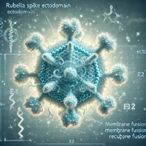
Molecular and Structural Insights into the E1-E2 Antigen
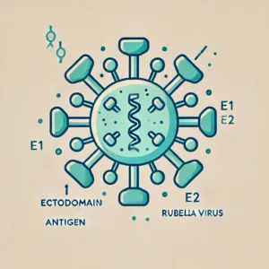
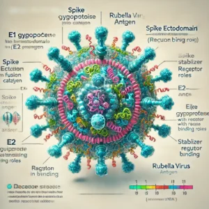
The spike ectodomain of RuV is primarily composed of two glycoproteins:
- E1 Glycoprotein: A class II fusion protein responsible for viral entry into host cells.
- E2 Glycoprotein: Stabilizes the E1 trimer and mediates receptor interactions critical for cell attachment.
E1 Glycoprotein: The Fusion Catalyst
The E1 glycoprotein drives the viral membrane fusion process, facilitating the injection of viral RNA into the host cytoplasm. Studies on the structural mechanisms of E1, published by NIH, demonstrate that specific regions within E1 are highly conserved, making it a prime target for neutralizing antibodies. The Protein Data Bank provides a repository of high-resolution structures that have been instrumental in understanding these functional domains.
E2 Glycoprotein: Enhancing Viral Stability
The E2 glycoprotein works in concert with E1, enhancing stability and ensuring proper viral attachment. Research from the CDC has highlighted the immunological relevance of E2 in triggering T-cell-mediated responses. Studies from Johns Hopkins University further underscore its role in modulating host immune evasion.
The combination of these glycoproteins in the E1-E2 ectodomain forms a critical target for vaccine development and diagnostics.
Role in Diagnostic Applications
The E1-E2 antigen is central to diagnostic tools for detecting RuV-specific antibodies. Its recombinant form has enabled the development of high-sensitivity ELISA kits. These kits are approved by regulatory agencies such as the FDA and recommended by the World Health Organization (WHO) for confirming rubella immunity, especially in prenatal care.
Advanced Diagnostics
- ELISA Tests: Incorporation of recombinant E1-E2 enhances test accuracy, reducing false positives. These assays, as reported by Stanford University, offer unparalleled sensitivity in detecting IgG and IgM antibodies.
- PCR-Based Detection: Quantitative PCR assays targeting E1 gene sequences provide rapid and reliable rubella diagnosis. The National Institute of Allergy and Infectious Diseases (NIAID) highlights their application in outbreak scenarios.
- Point-of-Care Devices: Portable diagnostic tools utilizing E1-E2 antigens, supported by Harvard University, are being deployed in low-resource settings to improve early detection and intervention.
Significance in Vaccine Development
Rubella vaccines are a cornerstone of global immunization programs, reducing rubella incidence and preventing CRS. Traditional live-attenuated vaccines include the full viral particle, but there is growing interest in antigen-specific subunit vaccines based on the E1-E2 ectodomain.
Subunit Vaccines Using E1-E2
- Advantages: Subunit vaccines minimize the risks associated with live attenuated strains, especially in immunocompromised individuals. They also provide opportunities for targeting specific immune responses, as documented in research from Oxford University.
- Recombinant Platforms: Studies published by MIT demonstrate how recombinant E1-E2 antigens expressed in Escherichia coli or insect cell systems enhance vaccine immunogenicity.
Research from the NIH Vaccine Research Center is actively investigating these next-generation vaccines, which are poised to meet the goals of the Global Rubella Elimination Initiative led by WHO.
Therapeutic Potential and Monoclonal Antibodies
The development of therapeutic monoclonal antibodies targeting the E1-E2 ectodomain is another promising avenue. High-affinity monoclonal antibodies are being tested for their ability to neutralize RuV and prevent severe outcomes in CRS. Data from the Mayo Clinic show encouraging preclinical results.
Monoclonal Antibody Development
- Neutralizing E1 Antibodies: Studies hosted by PubMed indicate that E1-specific antibodies can block viral entry effectively.
- Combination Therapies: Combining E1 and E2 targeting antibodies is under investigation at Yale University.
Global Implications and Future Directions
Rubella continues to challenge public health systems, particularly in regions where vaccination rates remain low. The E1-E2 spike ectodomain is at the forefront of efforts to combat this disease through innovative diagnostics, vaccines, and therapeutics.
Future Research Directions
- Structural Studies: Further investigation into the E1-E2 interaction using cryo-electron microscopy, as detailed by Nature, will provide deeper insights into therapeutic targeting.
- CRS Prevention: Collaborative research funded by Bill & Melinda Gates Foundation aims to optimize the use of E1-E2 antigens in vertical transmission prevention strategies.
- Immune Escape Mechanisms: Research at UC Berkeley is exploring mutations in the E1-E2 region that enable immune evasion, informing vaccine redesign.
Conclusion
The Rubella Spike Ectodomain (E1-E2) Antigen represents a crucial element in the fight against rubella. Its structural and immunological significance has enabled advancements in diagnostics, vaccine development, and therapeutic approaches. As global health organizations like the WHO and CDC work towards rubella elimination, the E1-E2 ectodomain remains central to these efforts.
For additional resources and research, visit trusted repositories like NIH, PubMed, and academic institutions such as Harvard and Stanford.
By leveraging the power of the E1-E2 antigen, the global medical community is poised to achieve significant milestones in rubella control and prevention.