
The AIF1/Iba1 (Microglia Marker) CF647 Conjugate Antibody is a specialized fluorescent antibody targeting Allograft Inflammatory Factor 1 (AIF1), also known as Iba1. This protein is a critical marker for microglia, the immune cells of the central nervous system (CNS), and plays a key role in neuroinflammation, neurodegeneration, and CNS homeostasis. The CF647 conjugation provides superior fluorescence properties for imaging and flow cytometry, making it an invaluable tool for advanced neurological research.
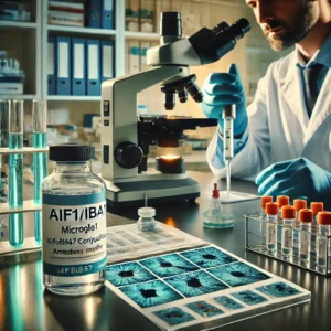
Overview of AIF1/Iba1
Key Features:
- Full Name: Allograft Inflammatory Factor 1 (AIF1), also known as Ionized Calcium-Binding Adaptor Molecule 1 (Iba1).
- Function:
- Regulates microglial activation.
- Involved in cytoskeletal reorganization.
- Facilitates phagocytosis and immune responses in the CNS.
- Clinical Significance:
- Highly expressed in activated microglia during neuroinflammatory and neurodegenerative conditions, such as Alzheimer’s disease, Parkinson’s disease, and multiple sclerosis.
For more information, visit NCBI Gene Database.
Features of the AIF1/Iba1 CF647 Conjugate Antibody
- Fluorescence Conjugation:
- Conjugated to CF647, a bright, photostable far-red fluorophore for superior imaging performance.
- Compatible with standard confocal and fluorescence microscopy filters.
- High Specificity:
- Targets AIF1/Iba1 with minimal cross-reactivity, ensuring precise identification of microglia.
- Versatility:
- Suitable for:
- Immunofluorescence (IF): Visualizes microglia in tissue sections.
- Flow Cytometry (FC): Quantifies microglial populations in cell suspensions.
- Suitable for:
- Species Reactivity:
- Validated for human, mouse, and rat samples.
- Ready-to-Use:
- Supplied at 0.1 mg/mL for immediate use in standard protocols.
Applications in Research
- Neuroinflammation Studies:
- Tracks microglial activation in response to injury or disease.
- Neurodegenerative Diseases:
- Examines microglial involvement in Alzheimer’s, Parkinson’s, and Huntington’s disease.
- Immune Responses in CNS:
- Studies the role of microglia in autoimmune disorders like multiple sclerosis.
- Drug Development:
- Evaluates therapeutic compounds targeting microglial activation and neuroinflammation.
Protocols for Using AIF1/Iba1 CF647 Antibody
Immunofluorescence (IF):
- Sample Preparation:
- Fix tissue sections with 4% paraformaldehyde.
- Permeabilize with 0.1% Triton X-100.
- Antibody Incubation:
- Dilute CF647 conjugated antibody (1:200–1:500 recommended).
- Incubate for 1 hour at room temperature in the dark.
- Imaging:
- Use a fluorescence microscope with appropriate filters for CF647.
Flow Cytometry (FC):
- Cell Preparation:
- Isolate microglial cells from CNS tissue using enzymatic digestion and gradient separation.
- Fix and permeabilize cells as needed.
- Staining:
- Incubate cells with the antibody diluted in buffer (1:50–1:200).
- Wash and resuspend in flow cytometry buffer.
- Detection:
- Use a flow cytometer with far-red laser compatibility.
Benefits of the AIF1/Iba1 CF647 Antibody
- High Sensitivity and Specificity:
- Accurately labels microglia in CNS tissues.
- Superior Fluorescence:
- CF647 offers brighter signals and reduced photobleaching compared to other dyes.
- Multifunctional:
- Suitable for imaging, flow cytometry, and co-localization studies.
- Reproducibility:
- Consistent results across various experimental setups.
Challenges and Future Directions
Challenges:
- Fluorophore Compatibility:
- Requires compatible microscopy and cytometry equipment.
- Optimization:
- Staining conditions may need adjustment for different sample types.
Future Innovations:
- Multiplex Antibody Panels:
- Combine with antibodies targeting other microglial markers for comprehensive profiling.
- Super-Resolution Imaging:
- Leverage CF647 for high-resolution studies of microglial dynamics.
- Therapeutic Applications:
- Utilize AIF1/Iba1 as a biomarker for microglial-targeted therapies.
Conclusion
The AIF1/Iba1 CF647 Conjugate Antibody is an advanced tool for exploring microglial biology and CNS immunity. Its high specificity, versatile applications, and superior fluorescence properties make it indispensable for cutting-edge research in neuroscience and immunology.
For further resources and technical support, visit: